Deposition Date
2008-01-30
Release Date
2008-12-30
Last Version Date
2024-05-01
Entry Detail
PDB ID:
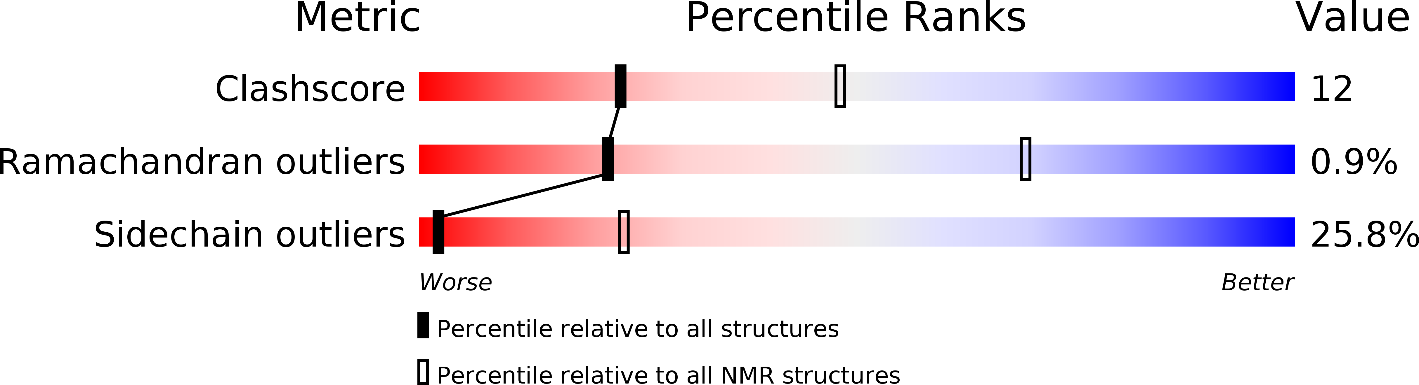
2RNN
Keywords:
Title:
Solution Structure of the N-terminal SAP Domain of SUMO E3 Ligases from Saccharomyces cerevisiae
Biological Source:
Source Organism(s):
Saccharomyces cerevisiae (Taxon ID: 4932)
Expression System(s):
Method Details:
Experimental Method:
Conformers Calculated:
100
Conformers Submitted:
20
Selection Criteria:
structures with the least restraint violations