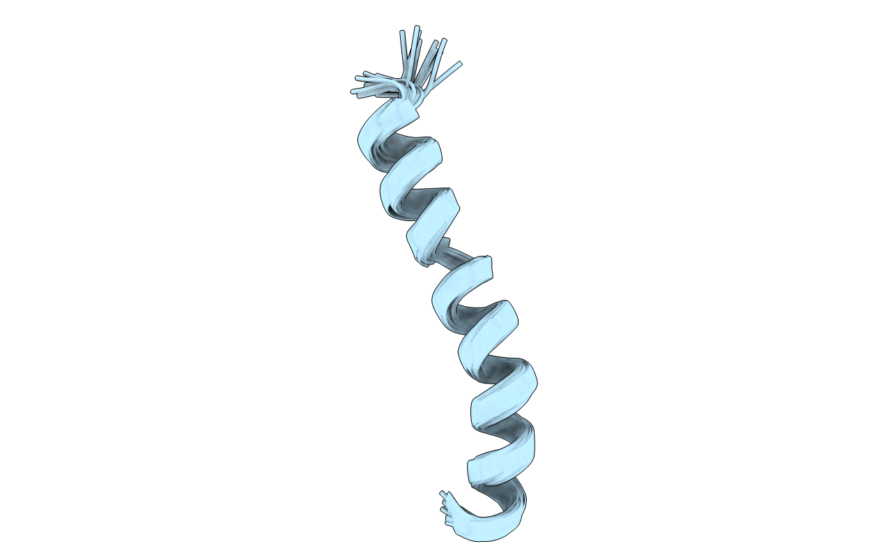
2RMI image
Deposition Date 2007-10-17
Release Date 2007-10-30
Last Version Date 2024-11-20
Entry Detail
PDB ID:
2RMI
Keywords:
Title:
3D NMR structure of astressin
Biological Source:
Source Organism:
Method Details:
Experimental Method:
Conformers Calculated:
100
Conformers Submitted:
20
Selection Criteria:
target function
Macromolecular Entities
Polymer Type:polypeptide(L)
Molecule:astressin
Chain IDs:A
Chain Length:30
Number of Molecules:1
Biological Source:
Modified Residue
Compound ID Chain ID Parent Comp ID Details 2D Image
NLE A LEU NORLEUCINE
Ligand Molecules
Primary Citation
Astressin-amide and astressin-acid are structurally different in dimethylsulfoxide
Biopolymers 87 196 205 (2007)
PMID: 17657708 DOI: 10.1002/bip.20818

Abstact

The C-terminally amidated CRF antagonist astressin binds to CRF-R1 or CRF-R2 receptors with low nanomolar affinity while the corresponding astressin-acid has >100 times less affinity. To understand the role of the amide group in binding, the conformations of astressin-amide and astressin-acid were studied in DMSO using NMR techniques. The 3D NMR structures show that the backbones of both analogs prefer an alpha-helical conformation, with a small kink around Gln(26). However, astressin-amide has a well-defined helical structure from Leu(27) to Ile(41) and a conformation very similar to the bioactive conformation reported by our group (Grace et al., Proc Natl Acad Sci USA 2007, 104, 4858-4863). In contrast, astressin-acid has an irregular helical conformation from Arg(35) onward, including a rearrangement of the side chains in that region. This structural difference highlights the crucial role of the C-terminal amidation for stabilization of astressin's bioactive conformation.

Legend

Protein

Chemical

Disease

Primary Citation of related structures