Deposition Date
2007-10-16
Release Date
2007-11-06
Last Version Date
2024-02-21
Entry Detail
PDB ID:
2RJZ
Keywords:
Title:
Crystal structure of the type 4 fimbrial biogenesis protein PilO from Pseudomonas aeruginosa
Biological Source:
Source Organism(s):
Pseudomonas aeruginosa (Taxon ID: 208964)
Expression System(s):
Method Details:
Experimental Method:
Resolution:
2.20 Å
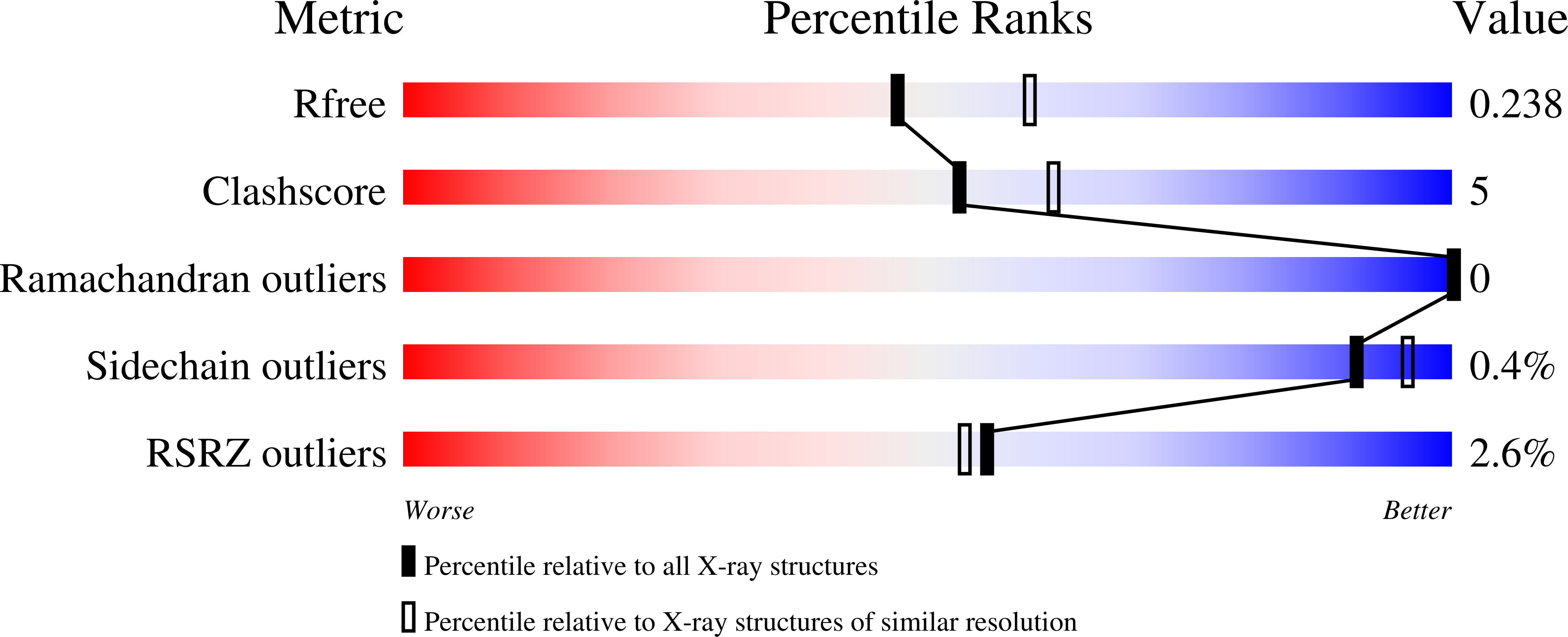
R-Value Free:
0.23
R-Value Work:
0.19
R-Value Observed:
0.19
Space Group:
P 64