Deposition Date
2007-10-15
Release Date
2008-01-22
Last Version Date
2024-02-21
Entry Detail
PDB ID:
2RJT
Keywords:
Title:
Crystal Structure Analysis of a Surface Entropy Reduction Mutant of S. pneumoniae FabF
Biological Source:
Source Organism(s):
Streptococcus pneumoniae (Taxon ID: 1313)
Expression System(s):
Method Details:
Experimental Method:
Resolution:
1.75 Å
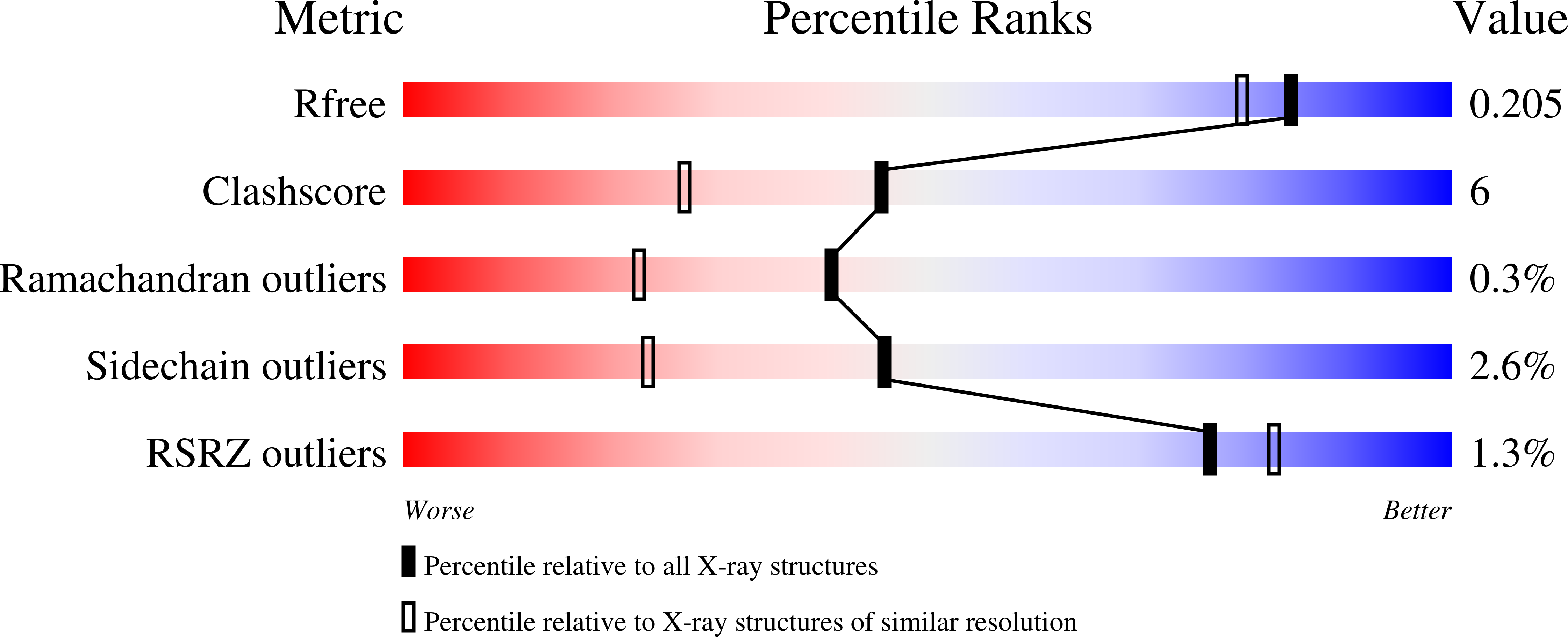
R-Value Free:
0.20
R-Value Work:
0.16
R-Value Observed:
0.16
Space Group:
C 2 2 21