2RIV image
Entry Detail
PDB ID:
2RIV
Title:
Crystal structure of the reactive loop cleaved human Thyroxine Binding Globulin
Biological Source:
Source Organism:
Host Organism:
PDB Version:
Deposition Date:
2007-10-12
Release Date:
2008-10-14
Method Details:
Experimental Method:
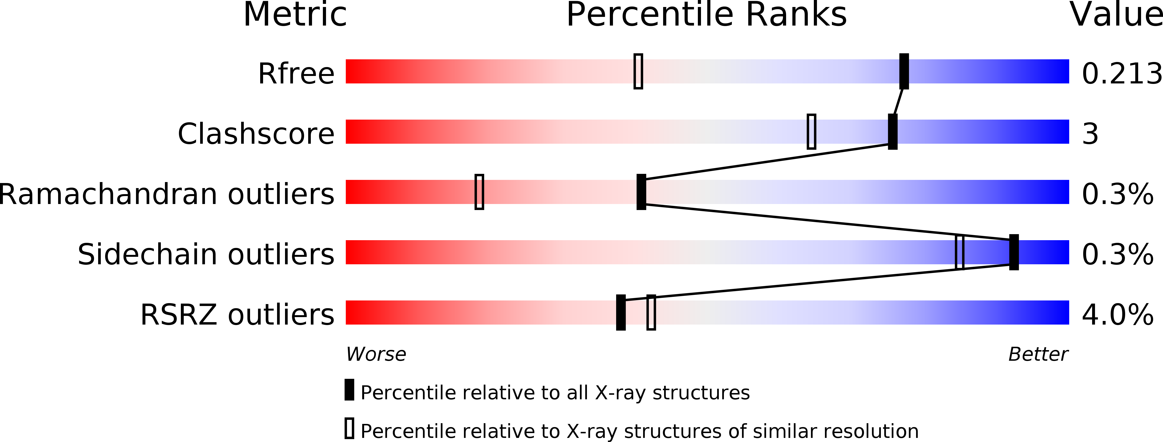
Resolution:
1.50 Å
R-Value Free:
0.21
R-Value Work:
0.18
R-Value Observed:
0.19
Space Group:
P 21 21 2
Macromolecular Entities
Polymer Type:polypeptide(L)
Description:Thyroxine-binding globulin
Chain IDs:A
Chain Length:343
Number of Molecules:1
Biological Source:Homo sapiens
Polymer Type:polypeptide(L)
Description:Thyroxine-binding globulin
Chain IDs:B
Chain Length:40
Number of Molecules:1
Biological Source:Homo sapiens
Primary Citation
Allosteric modulation of hormone release from thyroxine and corticosteroid-binding globulins
J.Biol.Chem. 286 16163 16173 (2011)
PMID: 21325280 DOI: 10.1074/jbc.M110.171082

Abstact

The release of hormones from thyroxine-binding globulin (TBG) and corticosteroid-binding globulin (CBG) is regulated by movement of the reactive center loop in and out of the β-sheet A of the molecule. To investigate how these changes are transmitted to the hormone-binding site, we developed a sensitive assay using a synthesized thyroxine fluorophore and solved the crystal structures of reactive loop cleaved TBG together with its complexes with thyroxine, the thyroxine fluorophores, furosemide, and mefenamic acid. Cleavage of the reactive loop results in its complete insertion into the β-sheet A and a substantial but incomplete decrease in binding affinity in both TBG and CBG. We show here that the direct interaction between residue Thr(342) of the reactive loop and Tyr(241) of the hormone binding site contributes to thyroxine binding and release following reactive loop insertion. However, a much larger effect occurs allosterically due to stretching of the connecting loop to the top of the D helix (hD), as confirmed in TBG with shortening of the loop by three residues, making it insensitive to the S-to-R transition. The transmission of the changes in the hD loop to the binding pocket is seen to involve coherent movements in the s2/3B loop linked to the hD loop by Lys(243), which is, in turn, linked to the s4/5B loop, flanking the thyroxine-binding site, by Arg(378). Overall, the coordinated movements of the reactive loop, hD, and the hormone binding site allow the allosteric regulation of hormone release, as with the modulation demonstrated here in response to changes in temperature.

Legend

Protein

Chemical

Disease

Primary Citation of related structures