2RHW image
Deposition Date 2007-10-09
Release Date 2007-11-13
Last Version Date 2024-02-21
Entry Detail
PDB ID:
2RHW
Keywords:
Title:
Crystal Structure of the S112A mutant of a C-C hydrolase, BphD from Burkholderia xenovorans LB400, in complex with 3,10-Di-Fluoro HOPDA
Biological Source:
Source Organism:
Method Details:
Experimental Method:
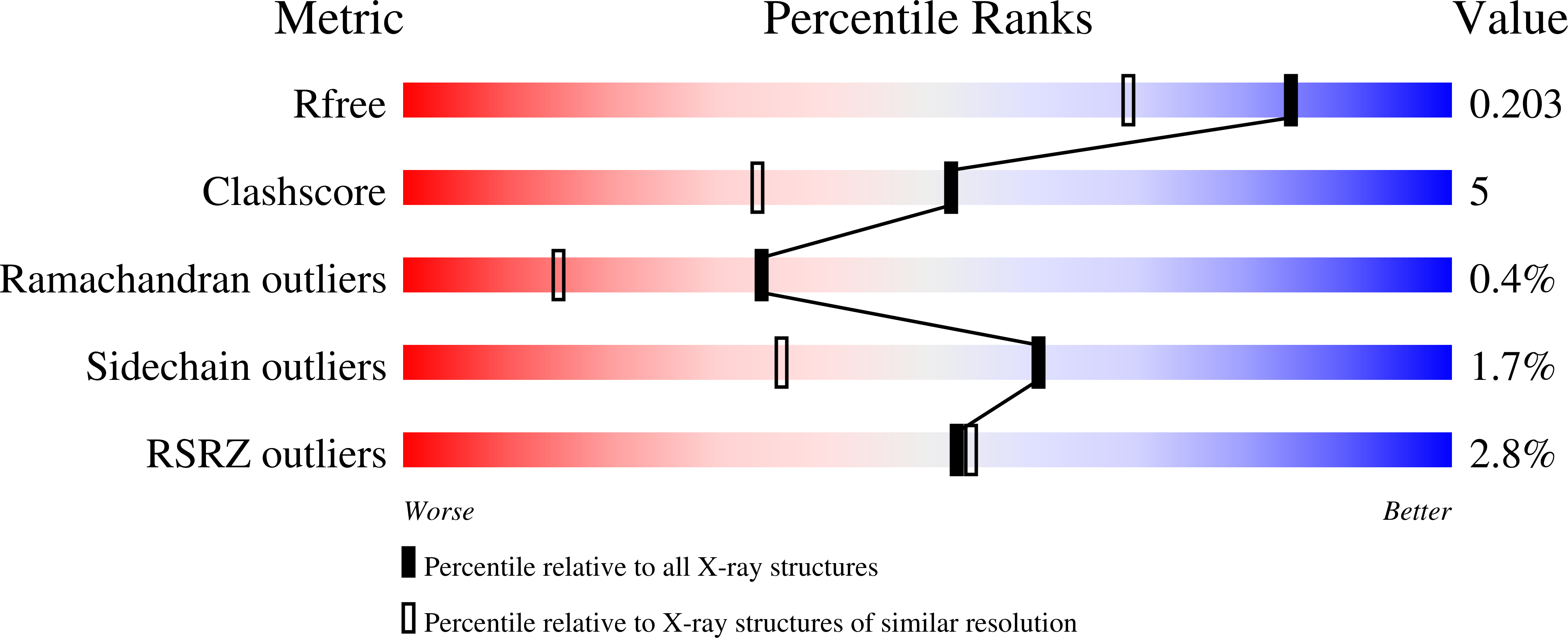
Resolution:
1.57 Å
R-Value Free:
0.20
R-Value Work:
0.17
R-Value Observed:
0.17
Space Group:
I 41 2 2
Macromolecular Entities
Polymer Type:polypeptide(L)
Molecule:2-hydroxy-6-oxo-6-phenylhexa-2,4-dienoate hydrolase
Gene (Uniprot):bphD
Mutations:S112A
Chain IDs:A
Chain Length:283
Number of Molecules:1
Biological Source:Burkholderia xenovorans
Primary Citation
The Molecular Basis for Inhibition of BphD, a C-C Bond Hydrolase Involved in Polychlorinated Biphenyls Degradation: LARGE 3-SUBSTITUENTS PREVENT TAUTOMERIZATION.
J.Biol.Chem. 282 36377 36385 (2007)
PMID: 17932031 DOI: 10.1074/jbc.M707035200

Abstact

The microbial degradation of polychlorinated biphenyls (PCBs) by the biphenyl catabolic (Bph) pathway is limited in part by the pathway's fourth enzyme, BphD. BphD catalyzes an unusual carbon-carbon bond hydrolysis of 2-hydroxy-6-oxo-6-phenylhexa-2,4-dienoic acid (HOPDA), in which the substrate is subject to histidine-mediated enol-keto tautomerization prior to hydrolysis. Chlorinated HOPDAs such as 3-Cl HOPDA inhibit BphD. Here we report that BphD preferentially hydrolyzed a series of 3-substituted HOPDAs in the order H>F>Cl>Me, suggesting that catalysis is affected by steric, not electronic, determinants. Transient state kinetic studies performed using wild-type BphD and the hydrolysis-defective S112A variant indicated that large 3-substituents inhibited His-265-catalyzed tautomerization by 5 orders of magnitude. Structural analyses of S112A.3-Cl HOPDA and S112A.3,10-diF HOPDA complexes revealed a non-productive binding mode in which the plane defined by the carbon atoms of the dienoate moiety of HOPDA is nearly orthogonal to that of the proposed keto tautomer observed in the S112A.HOPDA complex. Moreover, in the 3-Cl HOPDA complex, the 2-hydroxo group is moved by 3.6 A from its position near the catalytic His-265 to hydrogen bond with Arg-190 and access of His-265 is blocked by the 3-Cl substituent. Nonproductive binding may be stabilized by interactions involving the 3-substituent with non-polar side chains. Solvent molecules have poor access to C6 in the S112A.3-Cl HOPDA structure, more consistent with hydrolysis occurring via an acyl-enzyme than a gem-diol intermediate. These results provide insight into engineering BphD for PCB degradation.

Legend

Protein

Chemical

Disease

Primary Citation of related structures