Deposition Date
2007-10-09
Release Date
2008-07-01
Last Version Date
2024-02-21
Entry Detail
PDB ID:
2RHK
Keywords:
Title:
Crystal structure of influenza A NS1A protein in complex with F2F3 fragment of human cellular factor CPSF30, Northeast Structural Genomics Targets OR8C and HR6309A
Biological Source:
Source Organism(s):
Influenza A Virus (Taxon ID: 11320)
Homo sapiens (Taxon ID: 9606)
Homo sapiens (Taxon ID: 9606)
Expression System(s):
Method Details:
Experimental Method:
Resolution:
1.95 Å
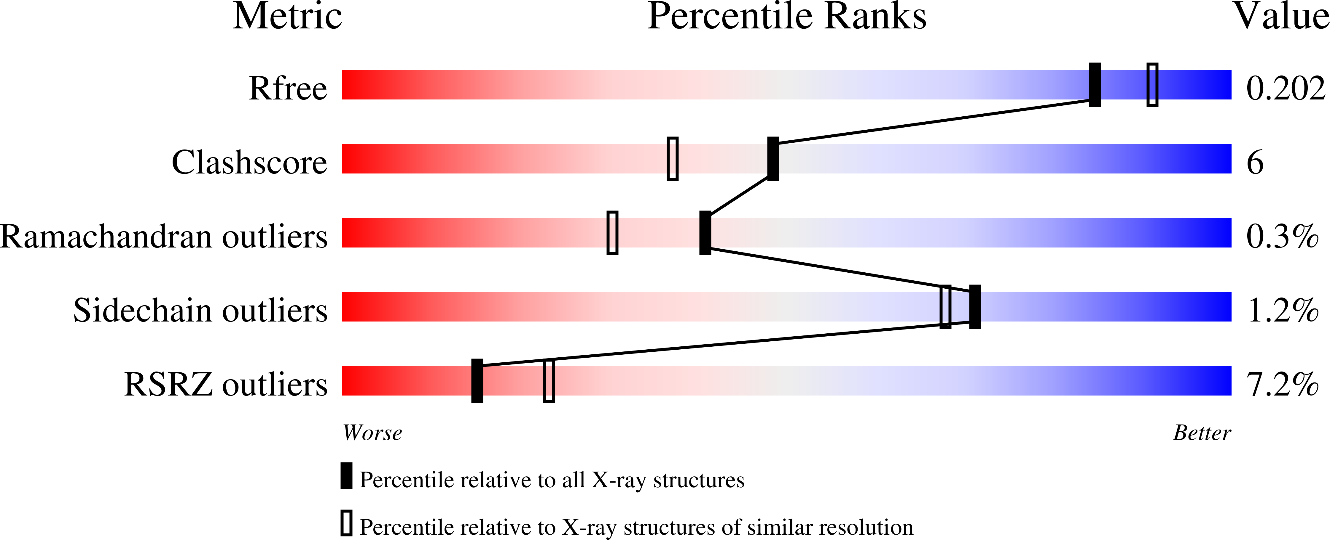
R-Value Free:
0.23
R-Value Work:
0.21
R-Value Observed:
0.21
Space Group:
P 41