
Deposition Date
2007-10-05
Release Date
2008-10-14
Last Version Date
2025-03-26
Entry Detail
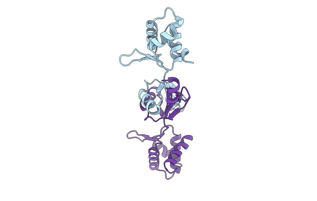
PDB ID:
2RGV
Keywords:
Title:
The crystal structure of PerR-Ox highlights 2-oxo-Histidine formation
Biological Source:
Source Organism(s):
Bacillus subtilis (Taxon ID: 1423)
Expression System(s):
Method Details:
Experimental Method:
Resolution:
2.05 Å
R-Value Free:
0.30
R-Value Work:
0.21
R-Value Observed:
0.21
Space Group:
P 1
