2RGI image
Deposition Date 2007-10-03
Release Date 2008-01-29
Last Version Date 2023-08-30
Entry Detail
PDB ID:
2RGI
Title:
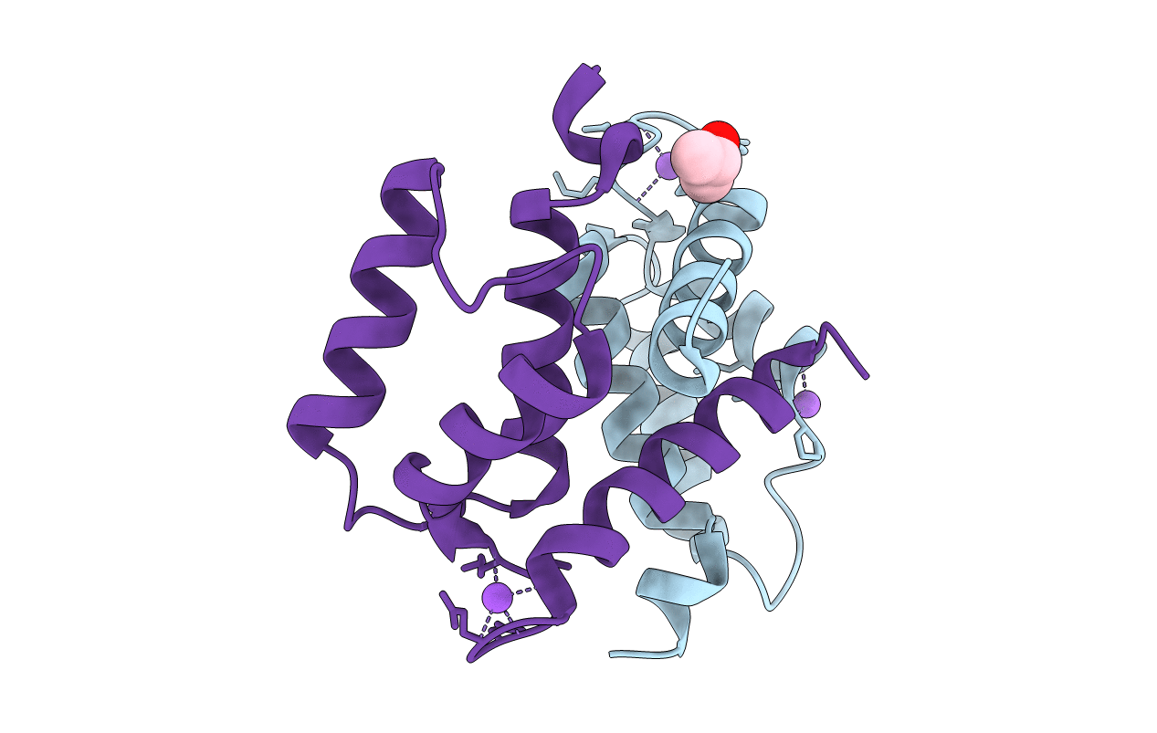
Crystal structure of Ca2+-free S100A2 at 1.6 A resolution
Biological Source:
Source Organism:
Homo sapiens (Taxon ID: 9606)
Method Details:
Experimental Method:
Resolution:
1.60 Å
R-Value Free:
0.22
R-Value Work:
0.18
R-Value Observed:
0.18
Space Group:
P 21 21 21
Macromolecular Entities
Structures with similar UniProt ID
Protein Blast
Polymer Type:polypeptide(L)
Molecule:Protein S100-A2
Gene (Uniprot):S100A2
Mutagens:C2S, C21S, C86S, C93S
Chain IDs:A, B
Chain Length:98
Number of Molecules:2
Biological Source:Homo sapiens
Primary Citation
Crystal structure of Ca2+ -free S100A2 at 1.6-A resolution.
J.Mol.Biol. 378 931 940 (2008)
PMID: 18394645 DOI: 10.1016/j.jmb.2008.03.019

Abstact

S100A2 is an EF hand-containing Ca(2+)-binding protein of the family of S100 proteins. The protein is localized exclusively in the nucleus and is involved in cell cycle regulation. It attracted most interest by its function as a tumor suppressor via p53 interaction. We determined the crystal structure of homodimeric S100A2 in the Ca(2+)-free state at 1.6-A resolution. The structure revealed structural differences between subunits A and B, especially in the conformation of a loop that connects the N- and C-terminal EF hands and represents a part of the target-binding site in S100 proteins. Analysis of the hydrogen bonding network and molecular dynamics calculations indicate that one of the two observed conformations is more stable. The structure revealed Na(+) bound to each N-terminal EF hand of both subunits coordinated by oxygen atoms of the backbone carbonyl and water molecules. Comparison with the structures of Ca(2+)-free S100A3 and S100A6 suggests that Na(+) might occupy the S100-specific EF hand in the Ca(2+)-free state.

Legend

Protein

Chemical

Disease

Primary Citation of related structures