Deposition Date
2007-10-01
Release Date
2008-01-15
Last Version Date
2024-10-30
Entry Detail
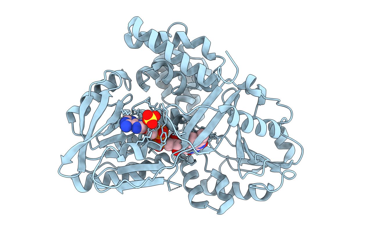
PDB ID:
2RGH
Keywords:
Title:
Structure of Alpha-Glycerophosphate Oxidase from Streptococcus sp.: A Template for the Mitochondrial Alpha-Glycerophosphate Dehydrogenase
Biological Source:
Source Organism(s):
Streptococcus sp. (Taxon ID: 1306)
Expression System(s):
Method Details:
Experimental Method:
Resolution:
2.30 Å
R-Value Free:
0.25
R-Value Work:
0.21
R-Value Observed:
0.21
Space Group:
P 21 21 2