2RFV image
Deposition Date 2007-10-02
Release Date 2008-08-19
Last Version Date 2023-11-15
Entry Detail
PDB ID:
2RFV
Keywords:
Title:
High resolution structure of L-methionine gamma-lyase from Citrobacter freundii
Biological Source:
Source Organism:
Host Organism:
Method Details:
Experimental Method:
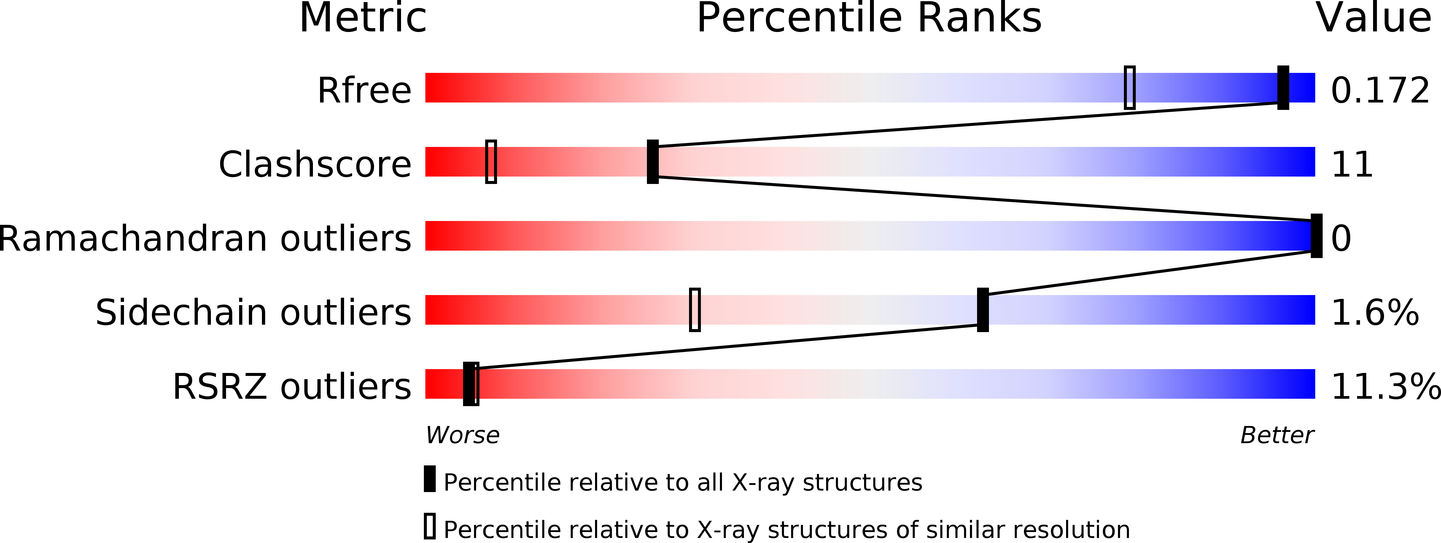
Resolution:
1.36 Å
R-Value Free:
0.17
R-Value Work:
0.15
Space Group:
I 2 2 2
Macromolecular Entities
Polymer Type:polypeptide(L)
Molecule:Methionine gamma-lyase
Gene (Uniprot):megL
Chain IDs:A
Chain Length:398
Number of Molecules:1
Biological Source:Citrobacter freundii
Modified Residue
Compound ID Chain ID Parent Comp ID Details 2D Image
CSO A CYS S-HYDROXYCYSTEINE
LLP A LYS ?
Ligand Molecules
Primary Citation
High-resolution structure of methionine gamma-lyase from Citrobacter freundii.
Acta Crystallogr.,Sect.D 64 211 218 (2008)
PMID: 18219122 DOI: 10.1107/S0907444907065390

Abstact

Pyridoxal 5'-phosphate-dependent methionine gamma-lyase (MGL) is involved in the metabolism of sulfur-containing amino acids. The enzyme is a promising target in some anaerobic pathogens and is effective in cancer-cell treatment. The structure of the MGL holoenzyme from Citrobacter freundii has previously been determined at 1.9 A resolution. By modification of the crystallization procedure, the previously determined structure of C. freundii MGL has been improved to 1.35 A resolution with R and R(free) values of 0.152 and 0.177, respectively. This high-resolution structure makes it possible to analyze the interactions between the monomers in detail and to reveal the structurally invariant regions that are responsible for monomer-monomer recognition during the formation of the active enzyme. Details of the mode of cofactor binding and of the flexible regions that may be involved in substrate recognition and binding are also described.

Legend

Protein

Chemical

Disease

Primary Citation of related structures