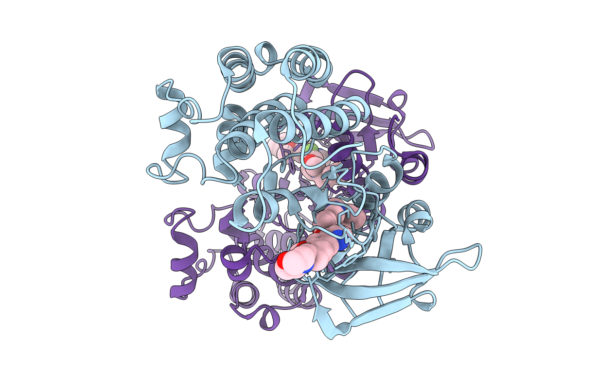
2RFN image
Entry Detail
PDB ID:
2RFN
Keywords:
Title:
x-ray structure of c-Met with inhibitor.
Biological Source:
Source Organism:
Host Organism:
PDB Version:
Deposition Date:
2007-10-01
Release Date:
2007-11-06
Method Details:
Experimental Method:
Resolution:
2.50 Å
R-Value Free:
0.29
R-Value Work:
0.23
R-Value Observed:
0.24
Space Group:
P 21 21 21
Macromolecular Entities
Polymer Type:polypeptide(L)
Description:Hepatocyte growth factor receptor
Chain IDs:A, B
Chain Length:310
Number of Molecules:2
Biological Source:Homo sapiens
Ligand Molecules
Primary Citation
c-Met inhibitors with novel binding mode show activity against several hereditary papillary renal cell carcinoma-related mutations.
J.Biol.Chem. 283 2675 2683 (2008)
PMID: 18055465 DOI: 10.1074/jbc.M705774200

Abstact

c-Met is a receptor tyrosine kinase often deregulated in human cancers, thus making it an attractive drug target. One mechanism by which c-Met deregulation leads to cancer is through gain-of-function mutations. Therefore, small molecules capable of targeting these mutations could offer therapeutic benefits for affected patients. SU11274 was recently described and reported to inhibit the activity of the wild-type and some mutant forms of c-Met, whereas other mutants are resistant to inhibition. We identified a novel series of c-Met small molecule inhibitors that are active against multiple mutants previously identified in hereditary papillary renal cell carcinoma patients. AM7 is active against wild-type c-Met as well as several mutants, inhibits c-Met-mediated signaling in MKN-45 and U-87 MG cells, and inhibits tumor growth in these two models grown as xenografts. The crystal structures of AM7 and SU11274 bound to unphosphorylated c-Met have been determined. The AM7 structure reveals a novel binding mode compared with other published c-Met inhibitors and SU11274. The molecule binds the kinase linker and then extends into a new hydrophobic binding site. This binding site is created by a significant movement of the C-helix and so represents an inactive conformation of the c-Met kinase. Thus, our results demonstrate that it is possible to identify and design inhibitors that will likely be active against mutants found in different cancers.

Legend

Protein

Chemical

Disease

Primary Citation of related structures