Deposition Date
2007-09-25
Release Date
2008-03-25
Last Version Date
2024-10-30
Entry Detail
PDB ID:
2RDZ
Keywords:
Title:
High Resolution Crystal Structure of the Escherichia coli Cytochrome c Nitrite Reductase.
Biological Source:
Source Organism(s):
Escherichia coli (Taxon ID: 562)
Method Details:
Experimental Method:
Resolution:
1.74 Å
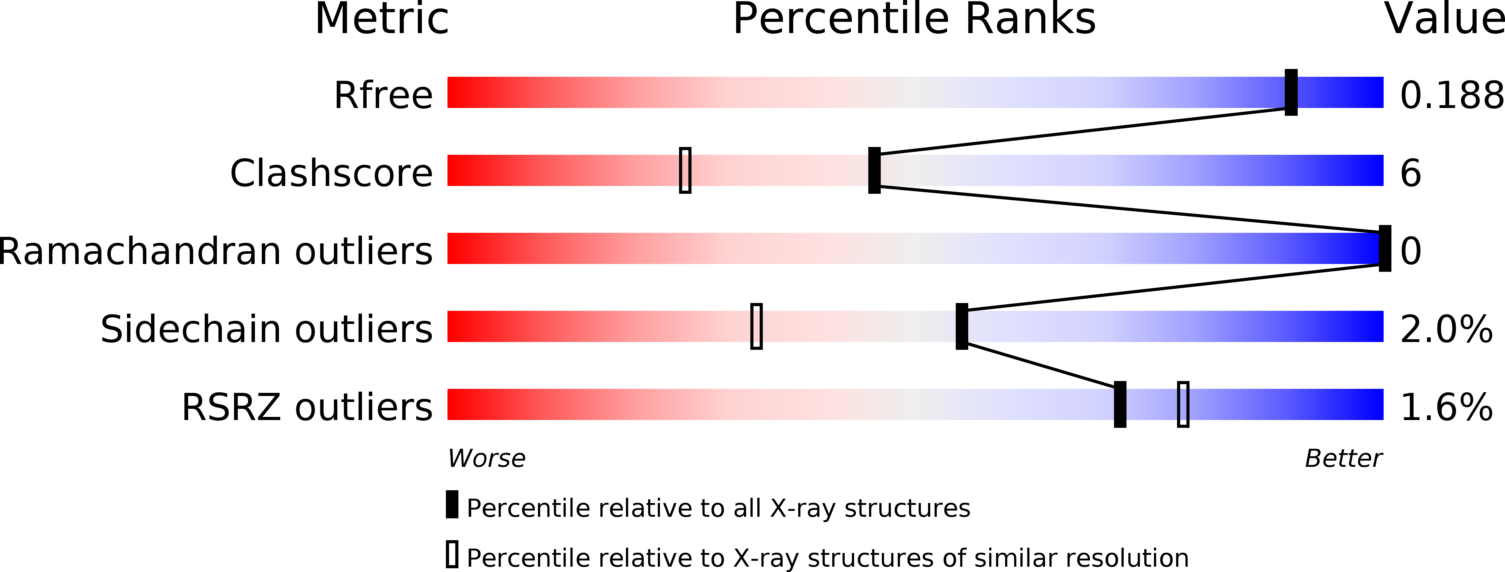
R-Value Free:
0.18
R-Value Work:
0.15
R-Value Observed:
0.15
Space Group:
P 1 21 1