
Deposition Date
2007-09-24
Release Date
2008-05-06
Last Version Date
2024-11-06
Entry Detail
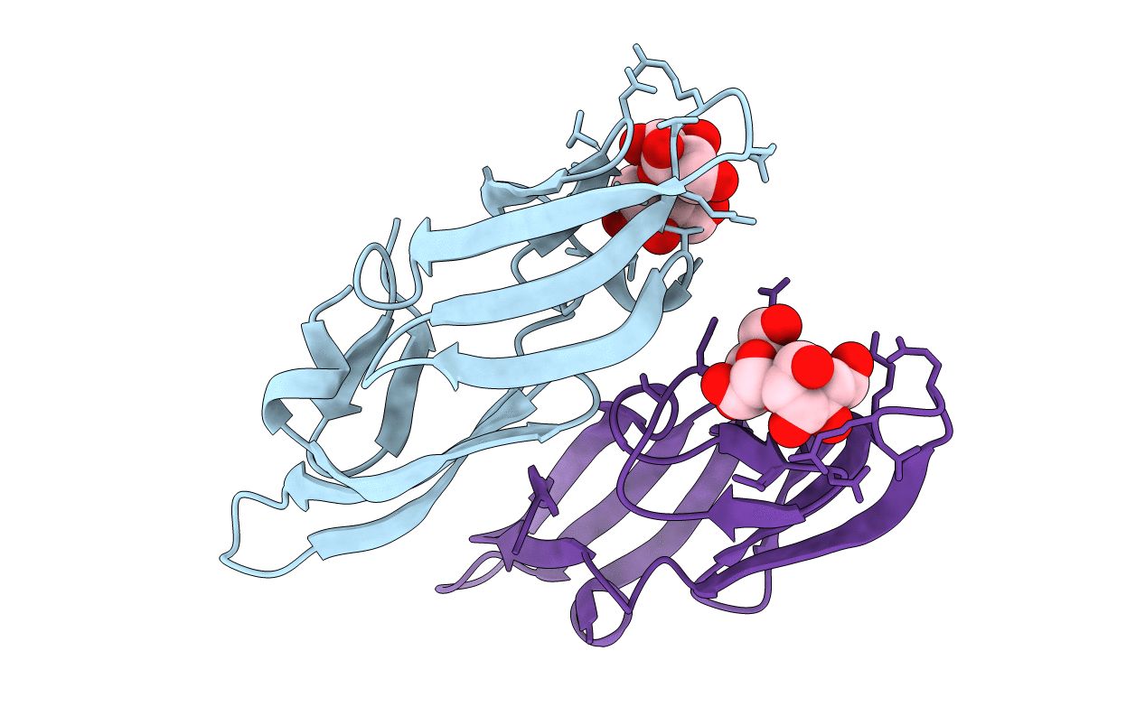
PDB ID:
2RDK
Keywords:
Title:
Five site mutated Cyanovirin-N with Mannose dimer bound
Biological Source:
Source Organism(s):
Nostoc ellipsosporum (Taxon ID: 45916)
Expression System(s):
Method Details:
Experimental Method:
Resolution:
1.35 Å
R-Value Free:
0.19
R-Value Work:
0.18
R-Value Observed:
0.18
Space Group:
P 1 21 1
