Deposition Date
2007-09-21
Release Date
2007-10-02
Last Version Date
2023-08-30
Entry Detail
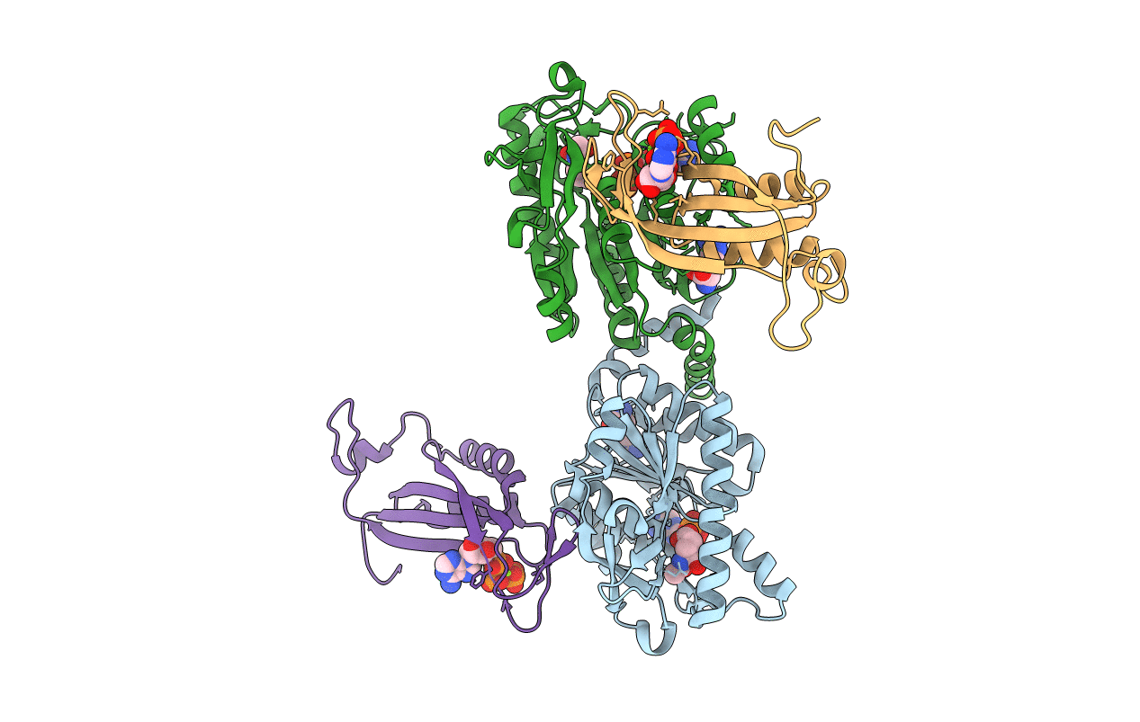
PDB ID:
2RD5
Keywords:
Title:
Structural basis for the regulation of N-acetylglutamate kinase by PII in Arabidopsis thaliana
Biological Source:
Source Organism(s):
Arabidopsis thaliana (Taxon ID: 3702)
Expression System(s):
Method Details:
Experimental Method:
Resolution:
2.51 Å
R-Value Free:
0.22
R-Value Work:
0.20
R-Value Observed:
0.20
Space Group:
P 21 3