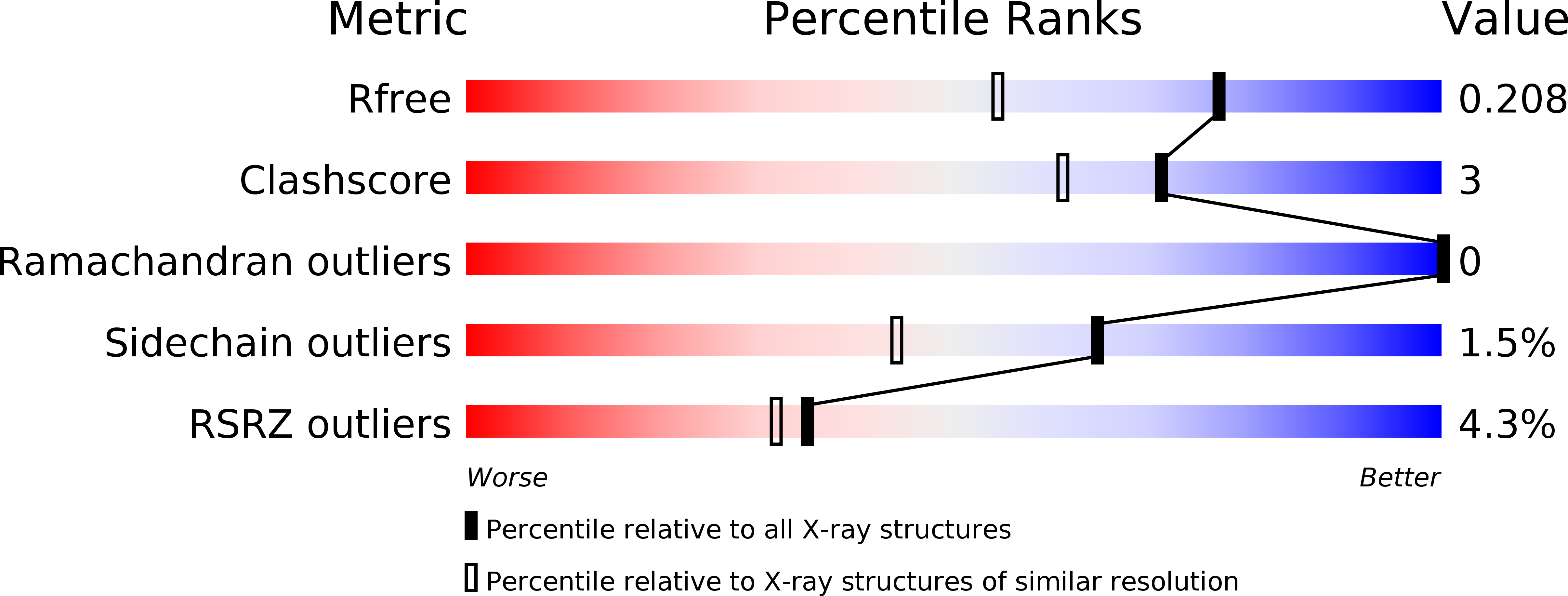
2RCV image
Deposition Date 2007-09-20
Release Date 2008-02-26
Last Version Date 2023-08-30
Entry Detail
PDB ID:
2RCV
Keywords:
Title:
Crystal structure of the Bacillus subtilis superoxide dismutase
Biological Source:
Source Organism:
Host Organism:
Method Details:
Experimental Method:
Resolution:
1.60 Å
R-Value Free:
0.23
R-Value Work:
0.21
R-Value Observed:
0.21
Space Group:
P 1
Macromolecular Entities
Structures with similar UniProt ID
Protein Blast
Polymer Type:polypeptide(L)
Molecule:Superoxide dismutase [Mn]
Gene (Uniprot):sodA
Chain IDs:A, B, C, D, E, F, G, H
Chain Length:202
Number of Molecules:8
Biological Source:Bacillus subtilis
Ligand Molecules
Primary Citation
Structure of Bacillus subtilis superoxide dismutase.
Acta Crystallogr.,Sect.F 63 1003 1007 (2007)
PMID: 18084079 DOI: 10.1107/S1744309107054127

Abstact

The sodA gene of Bacillus subtilis was expressed in Escherichia coli, purified and crystallized. The crystal structure of MnSOD was solved by molecular replacement with four dimers per asymmetric unit and refined to an R factor of 21.1% at 1.8 A resolution. The dimer structure is very similar to that of the related enzyme from B. anthracis. Larger structural differences were observed with the human MnSOD, which has one less helix in the helical domain and a longer loop between two beta-strands and also showed differences in three amino acids at the intersubunit interface in the dimer compared with the two bacterial MnSODs. These structural differences can be exploited in the design of drugs that selectively target the Bacillus enzymes.

Legend

Protein

Chemical

Disease

Primary Citation of related structures