Deposition Date
2007-09-17
Release Date
2007-11-06
Last Version Date
2024-10-30
Entry Detail
PDB ID:
2RAL
Keywords:
Title:
Crystal Structure Analysis of double cysteine mutant of S.epidermidis adhesin SdrG: Evidence for the Dock,Lock and Latch ligand binding mechanism
Biological Source:
Source Organism(s):
Staphylococcus epidermidis (Taxon ID: 1282)
Expression System(s):
Method Details:
Experimental Method:
Resolution:
2.80 Å
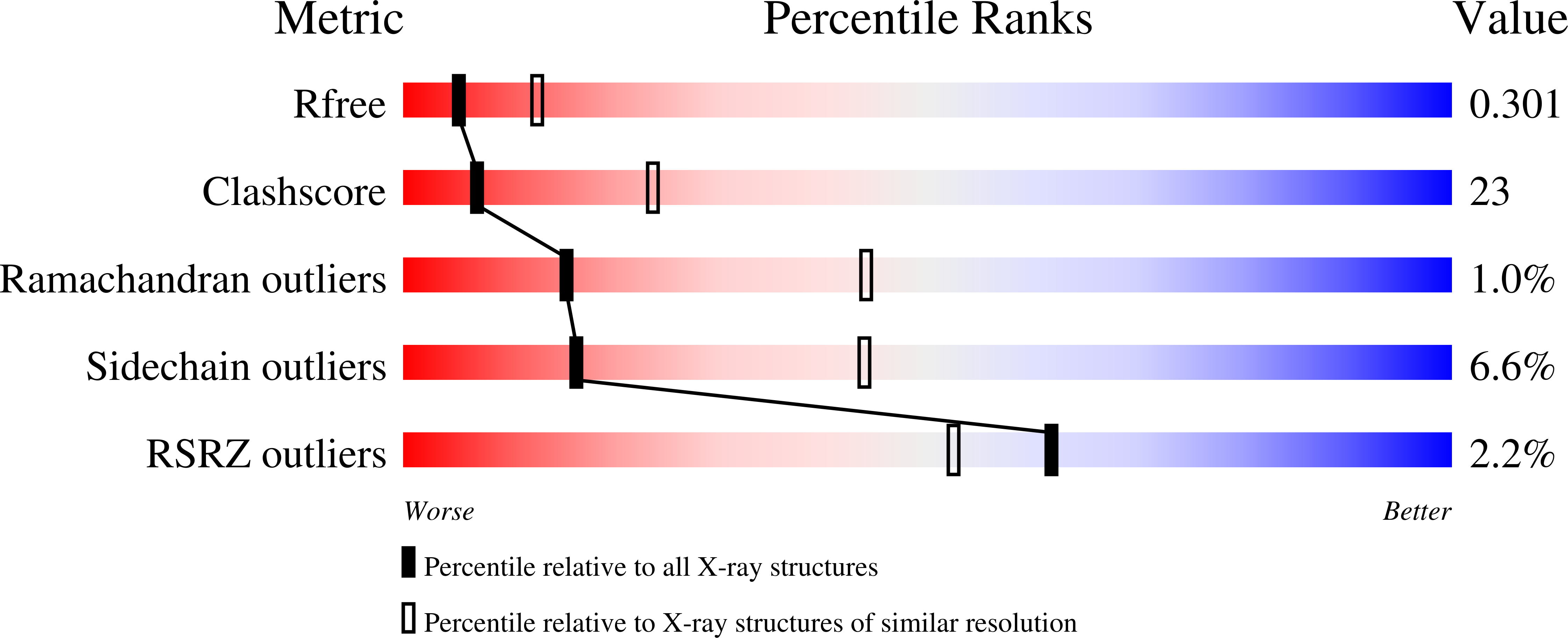
R-Value Free:
0.30
R-Value Work:
0.24
R-Value Observed:
0.24
Space Group:
P 21 21 21