2RA1 image
Deposition Date 2007-09-14
Release Date 2008-08-19
Last Version Date 2024-02-21
Entry Detail
PDB ID:
2RA1
Title:
Crystal structure of the N-terminal part of the bacterial S-layer protein SbsC
Biological Source:
Source Organism:
Host Organism:
Method Details:
Experimental Method:
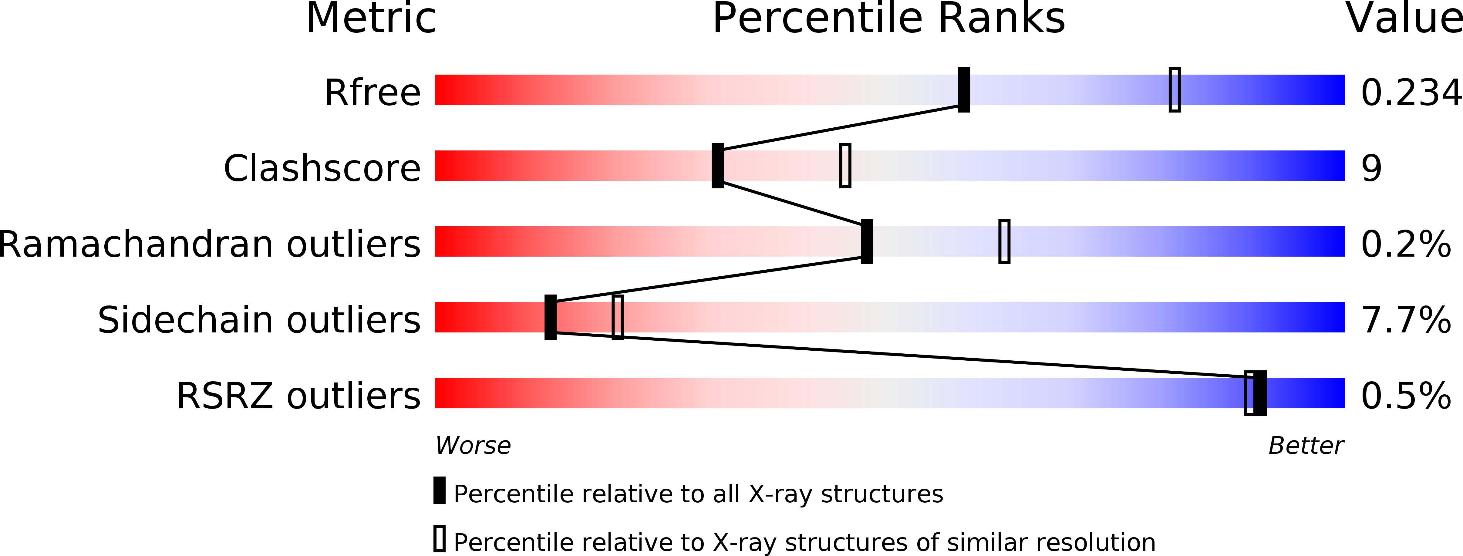
Resolution:
2.41 Å
R-Value Free:
0.24
R-Value Work:
0.17
Space Group:
P 1 21 1
Macromolecular Entities
Structures with similar UniProt ID
Protein Blast
Polymer Type:polypeptide(L)
Molecule:Surface layer protein
Gene (Uniprot):sbsC
Chain IDs:A
Chain Length:412
Number of Molecules:1
Biological Source:Geobacillus stearothermophilus
Primary Citation
The structure and binding behavior of the bacterial cell surface layer protein SbsC.
Structure 16 1226 1237 (2008)
PMID: 18682224 DOI: 10.1016/j.str.2008.05.012

Abstact

Surface layers (S-layers) comprise the outermost cell envelope component of most archaea and many bacteria. Here we present the structure of the bacterial S-layer protein SbsC from Geobacillus stearothermophilus, showing a very elongated and flexible molecule, with strong and specific binding to the secondary cell wall polymer (SCWP). The crystal structure of rSbsC((31-844)) revealed a novel fold, consisting of six separate domains, which are connected by short flexible linkers. The N-terminal domain exhibits positively charged residues regularly spaced along the putative ligand binding site matching the distance of the negative charges on the extended SCWP. Upon SCWP binding, a considerable stabilization of the N-terminal domain occurs. These findings provide insight into the processes of S-layer attachment to the underlying cell wall and self-assembly, and also accommodate the observed mechanical strength, the polarity of the S-layer, and the pronounced requirement for surface flexibility inherent to cell growth and division.

Legend

Protein

Chemical

Disease

Primary Citation of related structures