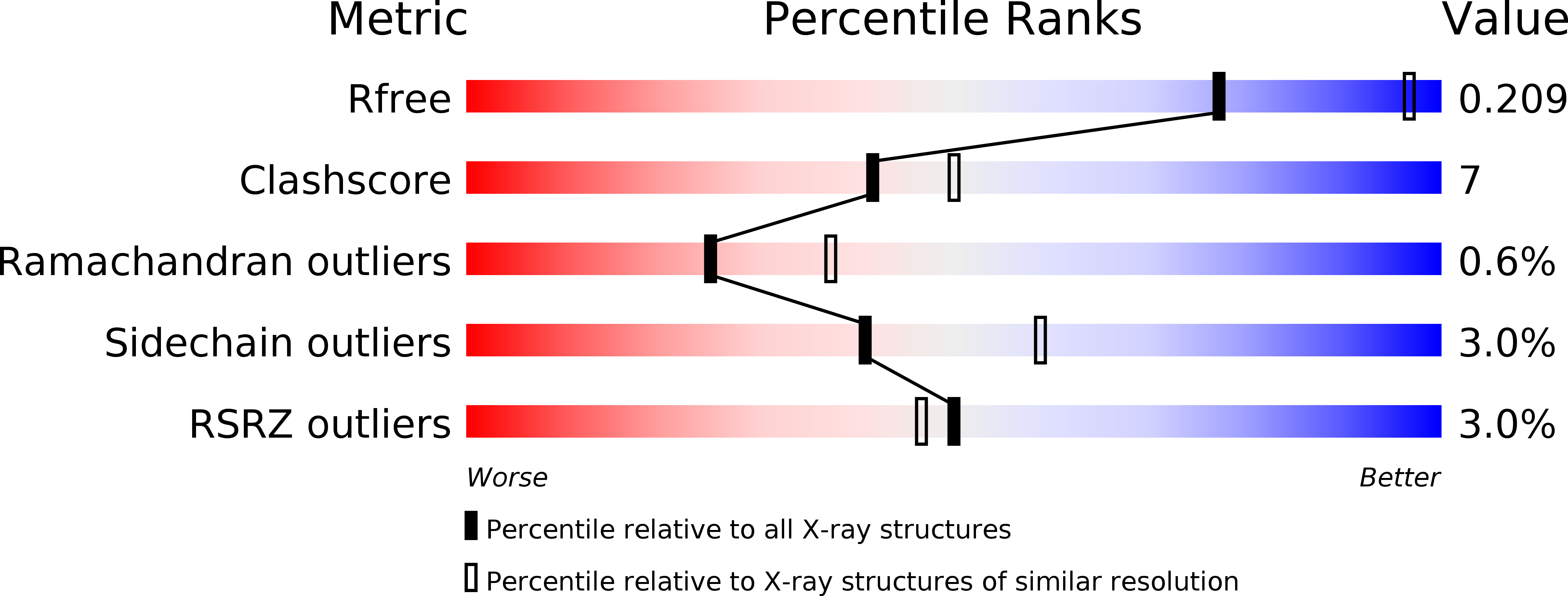
2R9Y image
Deposition Date 2007-09-14
Release Date 2007-12-18
Last Version Date 2023-10-25
Entry Detail
PDB ID:
2R9Y
Title:
Structure of antiplasmin
Biological Source:
Source Organism:
Mus musculus (Taxon ID: 10090)
Method Details:
Experimental Method:
Resolution:
2.65 Å
R-Value Free:
0.21
R-Value Work:
0.18
R-Value Observed:
0.18
Space Group:
P 65
Macromolecular Entities
Structures with similar UniProt ID
Protein Blast
Polymer Type:polypeptide(L)
Molecule:Alpha-2-antiplasmin
Gene (Uniprot):Serpinf2
Chain IDs:A
Chain Length:430
Number of Molecules:1
Biological Source:Mus musculus
Primary Citation
X-ray crystal structure of the fibrinolysis inhibitor {alpha}2-antiplasmin
Blood 111 2049 2052 (2008)
PMID: 18063751 DOI: 10.1182/blood-2007-09-114215

Abstact

The serpin alpha(2)-antiplasmin (SERPINF2) is the principal inhibitor of plasmin and inhibits fibrinolysis. Accordingly, alpha(2)-antiplasmin deficiency in humans results in uncontrolled fibrinolysis and a bleeding disorder. alpha(2)-antiplasmin is an unusual serpin, in that it contains extensive N- and C-terminal sequences flanking the serpin domain. The N-terminal sequence is crosslinked to fibrin by factor XIIIa, whereas the C-terminal region mediates the initial interaction with plasmin. To understand how this may happen, we have determined the 2.65A X-ray crystal structure of an N-terminal truncated murine alpha(2)-antiplasmin. The structure reveals that part of the C-terminal sequence is tightly associated with the body of the serpin. This would be anticipated to position the flexible plasmin-binding portion of the C-terminus in close proximity to the serpin Reactive Center Loop where it may act as a template to accelerate serpin/protease interactions.

Legend

Protein

Chemical

Disease

Primary Citation of related structures