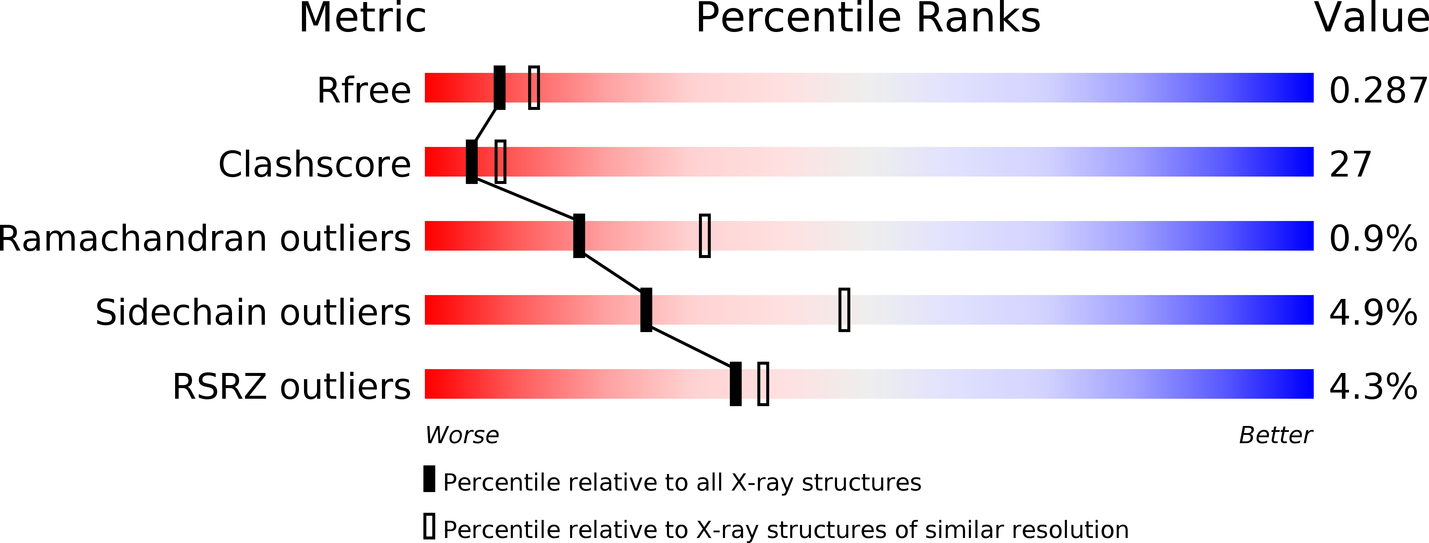
2R9A image
Deposition Date 2007-09-12
Release Date 2008-01-01
Last Version Date 2024-11-20
Entry Detail
PDB ID:
2R9A
Keywords:
Title:
Crystal structure of human XLF
Biological Source:
Source Organism:
Homo sapiens (Taxon ID: 9606)
Host Organism:
Method Details:
Experimental Method:
Resolution:
2.50 Å
R-Value Free:
0.28
R-Value Work:
0.25
R-Value Observed:
0.25
Space Group:
P 21 21 21
Macromolecular Entities
Polymer Type:polypeptide(L)
Molecule:Non-homologous end-joining factor 1
Gene (Uniprot):NHEJ1
Chain IDs:A, B
Chain Length:230
Number of Molecules:2
Biological Source:Homo sapiens
Modified Residue
Compound ID Chain ID Parent Comp ID Details 2D Image
MSE A MET SELENOMETHIONINE
Primary Citation
Crystal Structure of Human XLF: A Twist in Nonhomologous DNA End-Joining
Mol.Cell 28 1093 1101 (?)
PMID: 18158905 DOI: 10.1016/j.molcel.2007.10.024

Abstact

DNA double-strand breaks represent one of the most severe forms of DNA damage in mammalian cells. One pathway for repairing these breaks occurs via nonhomologous end-joining (NHEJ) and depends on XRCC4, LigaseIV, and Cernunnos, also called XLF. Although XLF stimulates XRCC4/LigaseIV to ligate mismatched and noncohesive DNA ends, the mechanistic basis for this function remains unclear. Here we report the structure of a partially functional 224 residue N-terminal fragment of human XLF. Despite only weak sequence similarity, XLF(1-170) shares structural homology with XRCC4(1-159). However, unlike the highly extended 130 A helical domain observed in XRCC4, XLF adopts a more compact, folded helical C-terminal region involving two turns and a twist, wrapping back to the structurally conserved N terminus. Mutational analysis of XLF and XRCC4 reveals a potential interaction interface, suggesting a mechanism for how XLF stimulates the ligation of mismatched ends.

Legend

Protein

Chemical

Disease

Primary Citation of related structures