
Deposition Date
2007-09-10
Release Date
2008-09-23
Last Version Date
2023-08-30
Entry Detail
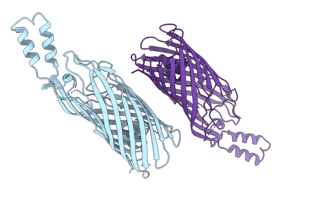
PDB ID:
2R88
Keywords:
Title:
Crystal structure of the long-chain fatty acid transporter FadL mutant delta S3 kink
Biological Source:
Source Organism(s):
Escherichia coli (Taxon ID: 562)
Expression System(s):
Method Details:
Experimental Method:
Resolution:
2.60 Å
R-Value Free:
0.29
R-Value Work:
0.26
R-Value Observed:
0.26
Space Group:
C 2 2 21
