2R83 image
Deposition Date 2007-09-10
Release Date 2008-02-12
Last Version Date 2023-08-30
Entry Detail
PDB ID:
2R83
Title:
Crystal structure analysis of human synaptotagmin 1 C2A-C2B
Biological Source:
Source Organism(s):
Homo sapiens (Taxon ID: 9606)
Expression System(s):
Method Details:
Experimental Method:
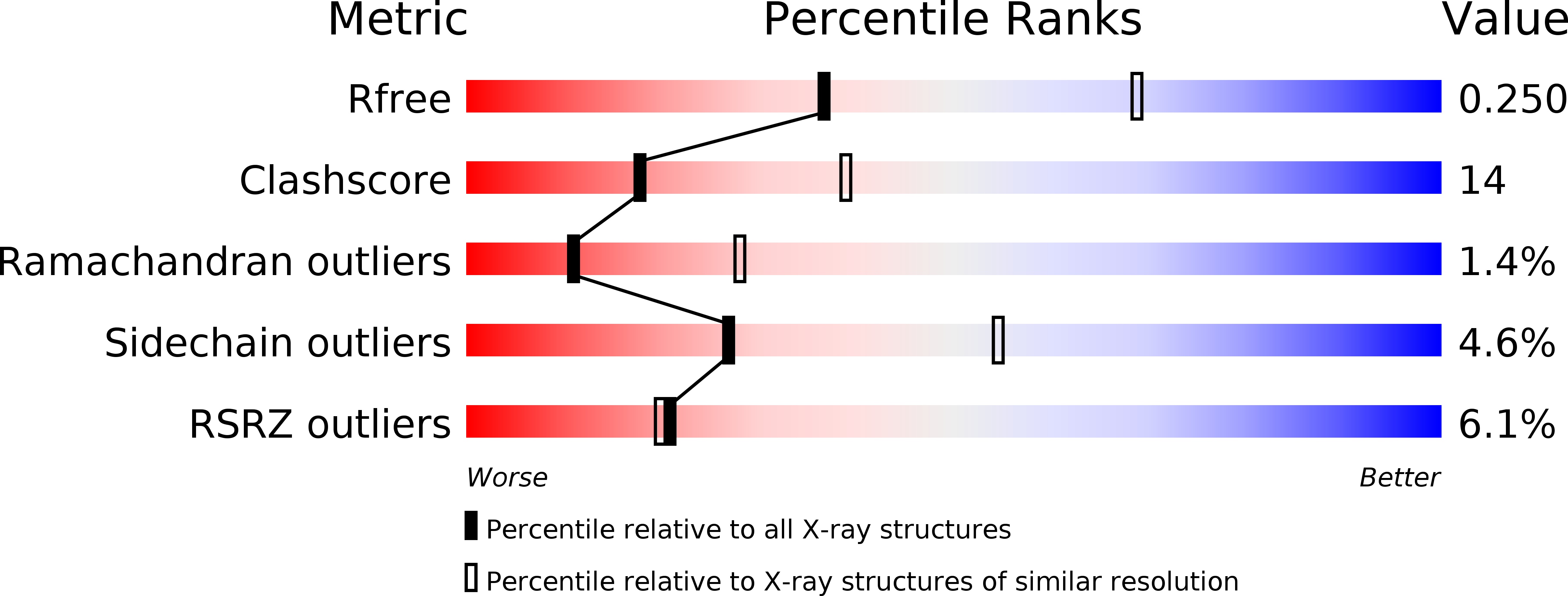
Resolution:
2.70 Å
R-Value Free:
0.25
R-Value Work:
0.23
R-Value Observed:
0.23
Space Group:
P 21 21 21
Macromolecular Entities
Structures with similar UniProt ID
Protein Blast
Polymer Type:polypeptide(L)
Molecule:Synaptotagmin-1
Gene (Uniprot):SYT1
Chain IDs:A, B
Chain Length:284
Number of Molecules:2
Biological Source:Homo sapiens
Ligand Molecules
Primary Citation
Structure of human synaptotagmin 1 C2AB in the absence of Ca2+ reveals a novel domain association.
Biochemistry 46 13041 13048 (2007)
PMID: 17956130 DOI: 10.1021/bi701651k

Abstact

Release of neurotransmitter from synaptic vesicles requires the Ca2+/phospholipid-binding protein synaptotagmin 1. There is considerable evidence that cooperation between the tandem C2 domains of synaptotagmin is a requirement of regulated exocytosis; however, high-resolution structural evidence for this interaction has been lacking. The 2.7 A crystal structure of the cytosolic domains of human synaptotagmin 1 in the absence of Ca2+ reveals a novel closed conformation of the protein. The shared interface between C2A and C2B is stabilized by a network of interactions between residues on the C-terminal alpha-helix of the C2B domain and residues on loops 1-3 of the Ca2+-binding region of C2A. These interactions alter the overall shape of the Ca2+-binding pocket of C2A, but not that of C2B. Thus, synaptotagmin 1 C2A-C2B may utilize a novel regulatory mechanism whereby one C2 domain could regulate the other until an appropriate triggering event decouples them.

Legend

Protein

Chemical

Disease

Primary Citation of related structures
Feedback Form
Name
Email
Institute
Feedback