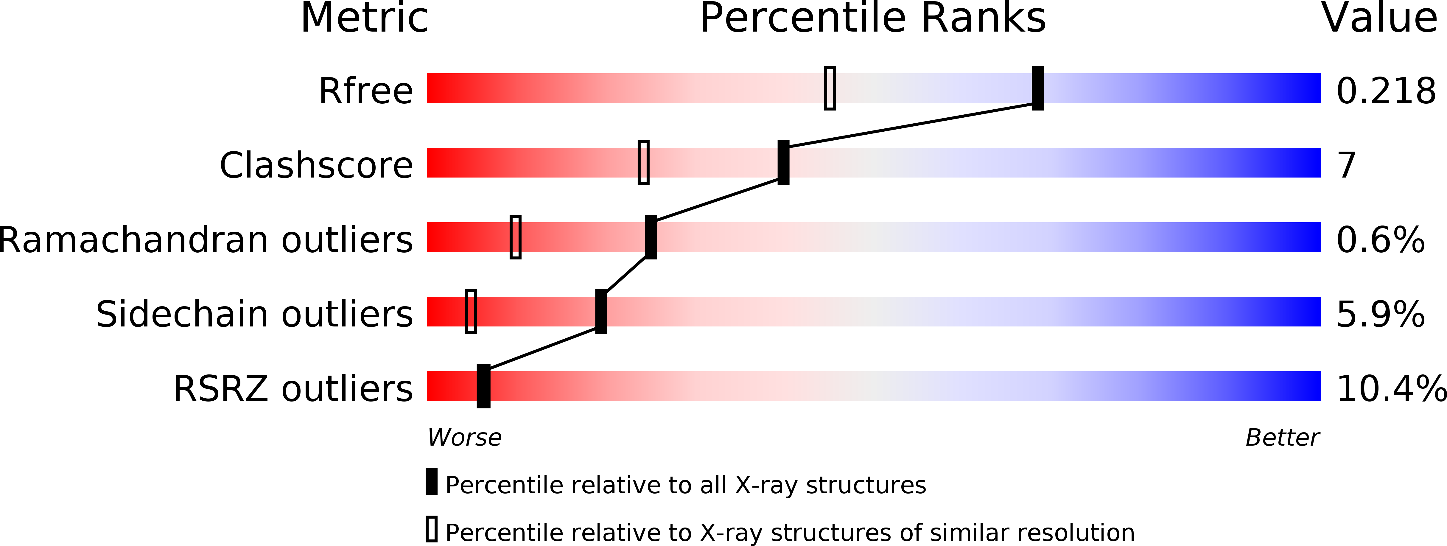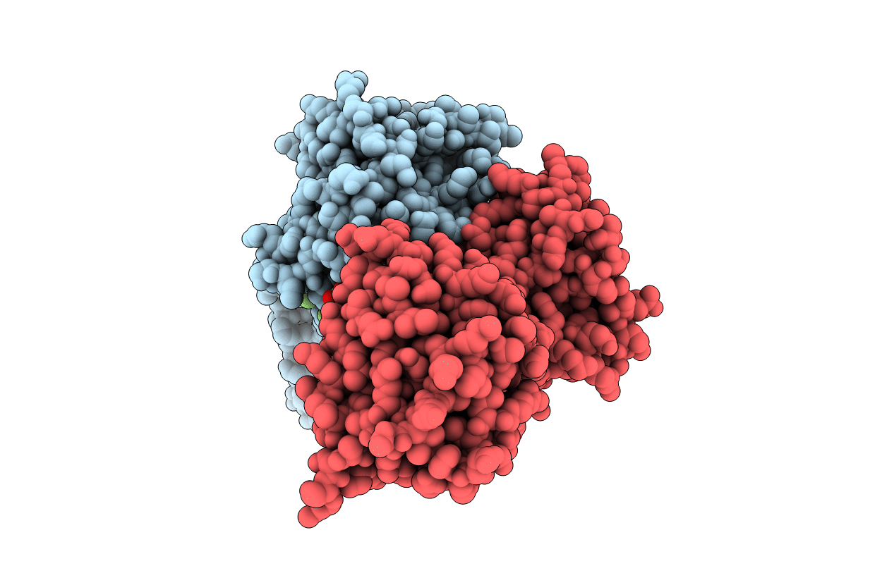
2R7G image
Deposition Date 2007-09-07
Release Date 2007-10-02
Last Version Date 2024-02-21
Entry Detail
PDB ID:
2R7G
Title:
Structure of the retinoblastoma protein pocket domain in complex with adenovirus E1A CR1 domain
Biological Source:
Source Organism:
Homo sapiens (Taxon ID: 9606)
Human adenovirus 5 (Taxon ID: 28285)
Method Details:
Experimental Method:
Resolution:
1.67 Å
R-Value Free:
0.21
R-Value Work:
0.19
R-Value Observed:
0.19
Space Group:
P 1
Macromolecular Entities
Structures with similar UniProt ID
Protein Blast
Polymer Type:polypeptide(L)
Molecule:Retinoblastoma-associated protein
Gene (Uniprot):RB1
Chain IDs:A, C
Chain Length:347
Number of Molecules:2
Biological Source:Homo sapiens
Structures with similar UniProt ID
Protein Blast
Polymer Type:polypeptide(L)
Molecule:Early E1A 32 kDa protein
Chain IDs:B, D, E
Chain Length:10
Number of Molecules:3
Biological Source:Human adenovirus 5
Ligand Molecules
Primary Citation
Structure of the retinoblastoma protein bound to adenovirus E1A reveals the molecular basis for viral oncoprotein inactivation of a tumor suppressor
Genes Dev. 21 2711 2716 (2007)
PMID: 17974914 DOI: 10.1101/gad.1590607

Abstact

The adenovirus (Ad) E1A (Ad-E1A) oncoprotein mediates cell transformation, in part, by displacing E2F transcription factors from the retinoblastoma protein (pRb) tumor suppressor. In this study we determined the crystal structure of the pRb pocket domain in complex with conserved region 1 (CR1) of Ad5-E1A. The structure and accompanying biochemical studies reveal that E1A-CR1 binds at the interface of the A and B cyclin folds of the pRb pocket domain, and that both E1A-CR1 and the E2F transactivation domain use similar conserved nonpolar residues to engage overlapping sites on pRb, implicating a novel molecular mechanism for pRb inactivation by a viral oncoprotein.

Legend

Protein

Chemical

Disease

Primary Citation of related structures