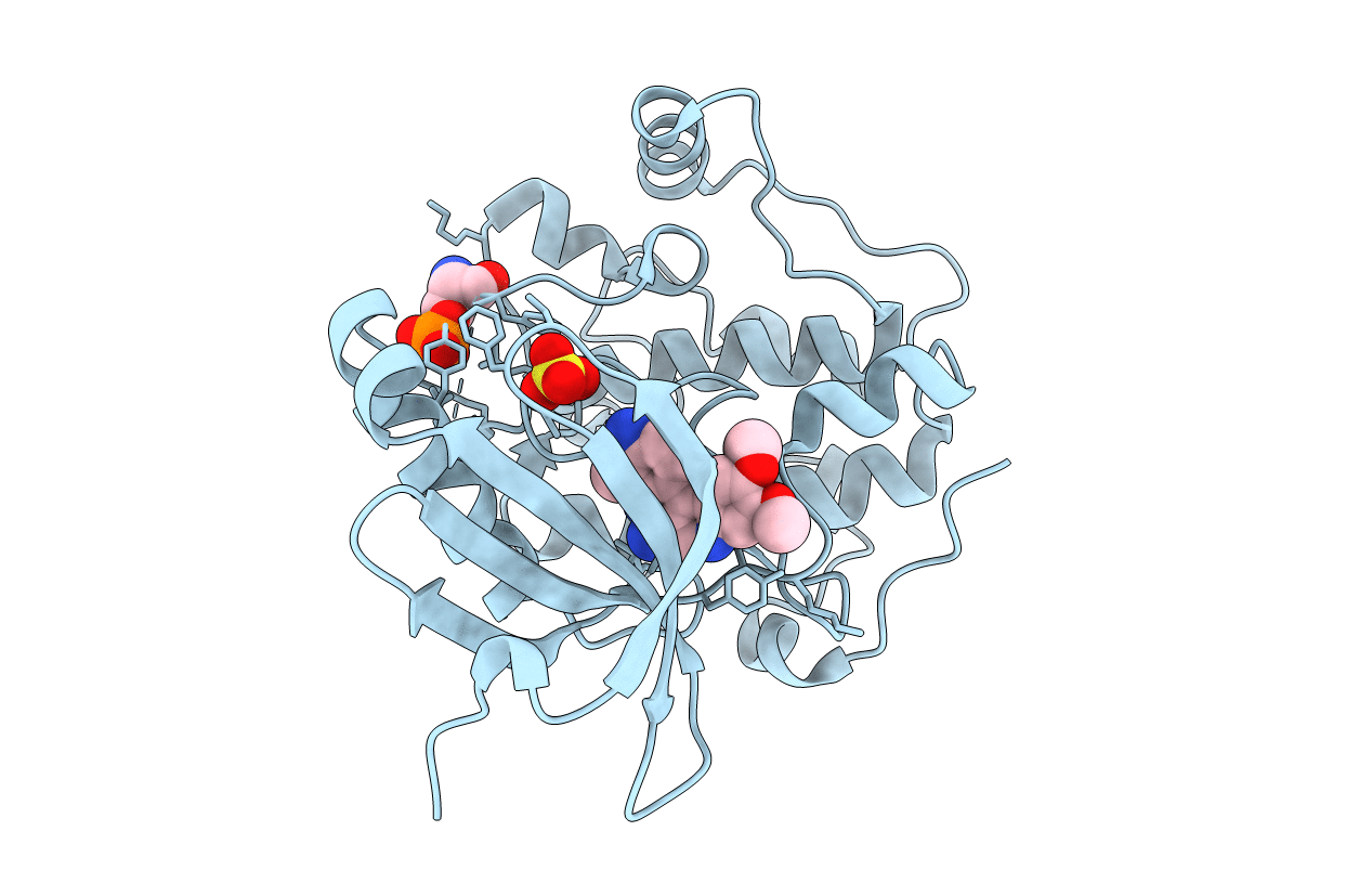
2R7B image
Deposition Date 2007-09-07
Release Date 2008-09-09
Last Version Date 2024-11-06
Entry Detail
PDB ID:
2R7B
Title:
Crystal Structure of the Phosphoinositide-dependent Kinase-1 (PDK-1)Catalytic Domain bound to a dibenzonaphthyridine inhibitor
Biological Source:
Source Organism:
Homo sapiens (Taxon ID: 9606)
Host Organism:
Method Details:
Experimental Method:
Resolution:
2.70 Å
R-Value Free:
0.24
R-Value Work:
0.22
Space Group:
P 32 2 1
Macromolecular Entities
Structures with similar UniProt ID
Protein Blast
Polymer Type:polypeptide(L)
Molecule:3-phosphoinositide-dependent protein kinase 1
Gene (Uniprot):PDPK1
Chain IDs:A
Chain Length:312
Number of Molecules:1
Biological Source:Homo sapiens
Modified Residue
Compound ID Chain ID Parent Comp ID Details 2D Image
SEP A SER modified residue
Primary Citation
Discovery of Dibenzo[c,f][2,7]naphthyridines as Potent and Selective 3-Phosphoinositide-Dependent Kinase-1 Inhibitors.
J.Med.Chem. 50 5547 5549 (2007)
PMID: 17941624 DOI: 10.1021/jm070851i

Abstact

With high-throughput screening, substituted dibenzo[c,f][2,7]naphthyridine 1 was identified as a novel potent and selective phosphoinositide-dependent kinase-1 (PDK-1) inhibitor. Various regions of the lead molecule were explored to understand the SAR requirement for this scaffold. The crystal structure of 1 with kinase domain of PDK-1 confirmed the binding in the active site. The key interaction of the molecule with the active site residues, observed SAR, and the biological profile are discussed in detail.

Legend

Protein

Chemical

Disease

Primary Citation of related structures