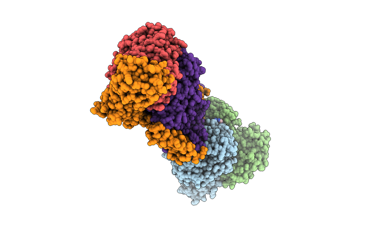
2R6G image
Deposition Date 2007-09-05
Release Date 2007-11-27
Last Version Date 2023-08-30
Entry Detail
PDB ID:
2R6G
Title:
The Crystal Structure of the E. coli Maltose Transporter
Biological Source:
Source Organism:
Host Organism:
Method Details:
Experimental Method:
Resolution:
2.80 Å
R-Value Free:
0.27
R-Value Work:
0.24
R-Value Observed:
0.24
Space Group:
P 1
Macromolecular Entities
Polymer Type:polypeptide(L)
Molecule:Maltose/maltodextrin import ATP-binding protein malK
Mutations:E159Q
Chain IDs:A, B
Chain Length:381
Number of Molecules:2
Biological Source:Escherichia coli
Polymer Type:polypeptide(L)
Molecule:Maltose-binding periplasmic protein
Gene (Uniprot):malE
Chain IDs:C (auth: E)
Chain Length:370
Number of Molecules:1
Biological Source:Escherichia coli
Polymer Type:polypeptide(L)
Molecule:Maltose transport system permease protein malF
Gene (Uniprot):malF
Chain IDs:D (auth: F)
Chain Length:514
Number of Molecules:1
Biological Source:Escherichia coli
Polymer Type:polypeptide(L)
Molecule:Maltose transport system permease protein malG
Gene (Uniprot):malG
Chain IDs:E (auth: G)
Chain Length:296
Number of Molecules:1
Biological Source:Escherichia coli
Ligand Molecules
Peptide-like Molecules
PRD_900001
Primary Citation
Crystal structure of a catalytic intermediate of the maltose transporter.
Nature 450 515 521 (2007)
PMID: 18033289 DOI: 10.1038/nature06264

Abstact

The maltose uptake system of Escherichia coli is a well-characterized member of the ATP-binding cassette transporter superfamily. Here we present the 2.8-A crystal structure of the intact maltose transporter in complex with the maltose-binding protein, maltose and ATP. This structure, stabilized by a mutation that prevents ATP hydrolysis, captures the ATP-binding cassette dimer in a closed, ATP-bound conformation. Maltose is occluded within a solvent-filled cavity at the interface of the two transmembrane subunits, about halfway into the lipid bilayer. The binding protein docks onto the entrance of the cavity in an open conformation and serves as a cap to ensure unidirectional translocation of the sugar molecule. These results provide direct evidence for a concerted mechanism of transport in which solute is transferred from the binding protein to the transmembrane subunits when the cassette dimer closes to hydrolyse ATP.

Legend

Protein

Chemical

Disease

Primary Citation of related structures