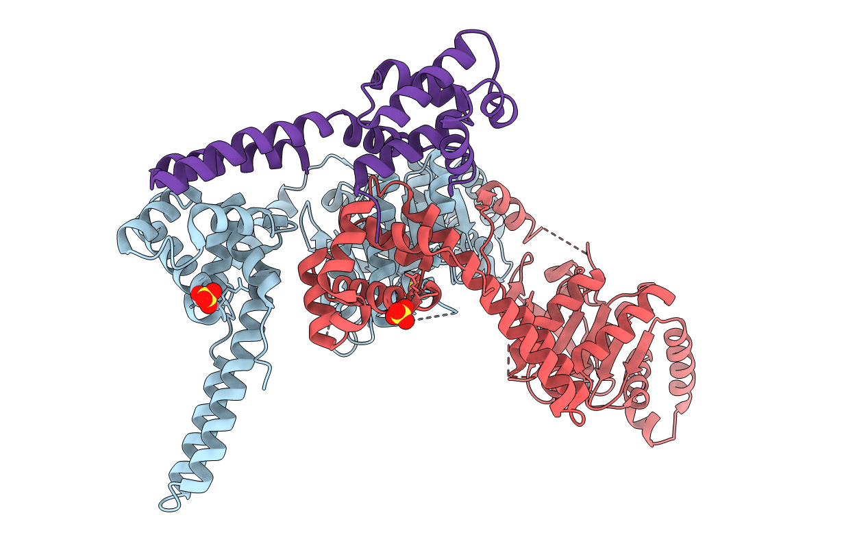
2R6A image
Deposition Date 2007-09-05
Release Date 2007-11-06
Last Version Date 2024-11-13
Entry Detail
PDB ID:
2R6A
Keywords:
Title:
Crystal Form BH1
Biological Source:
Source Organism:
Method Details:
Experimental Method:
Resolution:
2.90 Å
R-Value Free:
0.29
R-Value Work:
0.25
R-Value Observed:
0.26
Space Group:
P 3 2 1
Macromolecular Entities
Structures with similar UniProt ID
Protein Blast
Polymer Type:polypeptide(L)
Molecule:Replicative helicase
Gene (Uniprot):dnaB
Chain IDs:A, B
Chain Length:454
Number of Molecules:2
Biological Source:Geobacillus stearothermophilus
Structures with similar UniProt ID
Protein Blast
Polymer Type:polypeptide(L)
Molecule:DnaG Primase, Helicase Binding Domain
Gene (Uniprot):dnaG
Chain IDs:C
Chain Length:143
Number of Molecules:1
Biological Source:Geobacillus stearothermophilus
Modified Residue
Compound ID Chain ID Parent Comp ID Details 2D Image
MSE C MET SELENOMETHIONINE
Ligand Molecules
Primary Citation
Structure of hexameric DnaB helicase and its complex with a domain of DnaG primase
Science 318 459 463 (2007)
PMID: 17947583 DOI: 10.1126/science.1147353

Abstact

The complex between the DnaB helicase and the DnaG primase unwinds duplex DNA at the eubacterial replication fork and synthesizes the Okazaki RNA primers. The crystal structures of hexameric DnaB and its complex with the helicase binding domain (HBD) of DnaG reveal that within the hexamer the two domains of DnaB pack with strikingly different symmetries to form a distinct two-layered ring structure. Each of three bound HBDs stabilizes the DnaB hexamer in a conformation that may increase its processivity. Three positive, conserved electrostatic patches on the N-terminal domain of DnaB may also serve as a binding site for DNA and thereby guide the DNA to a DnaG active site.

Legend

Protein

Chemical

Disease

Primary Citation of related structures