Deposition Date
2007-09-02
Release Date
2007-11-27
Last Version Date
2023-10-25
Entry Detail
PDB ID:
2R4X
Keywords:
Title:
Ligand Migration and Binding in The Dimeric Hemoglobin of Scapharca Inaequivalvis: H69V/I114M co complex
Biological Source:
Source Organism(s):
Scapharca inaequivalvis (Taxon ID: 6561)
Expression System(s):
Method Details:
Experimental Method:
Resolution:
2.10 Å
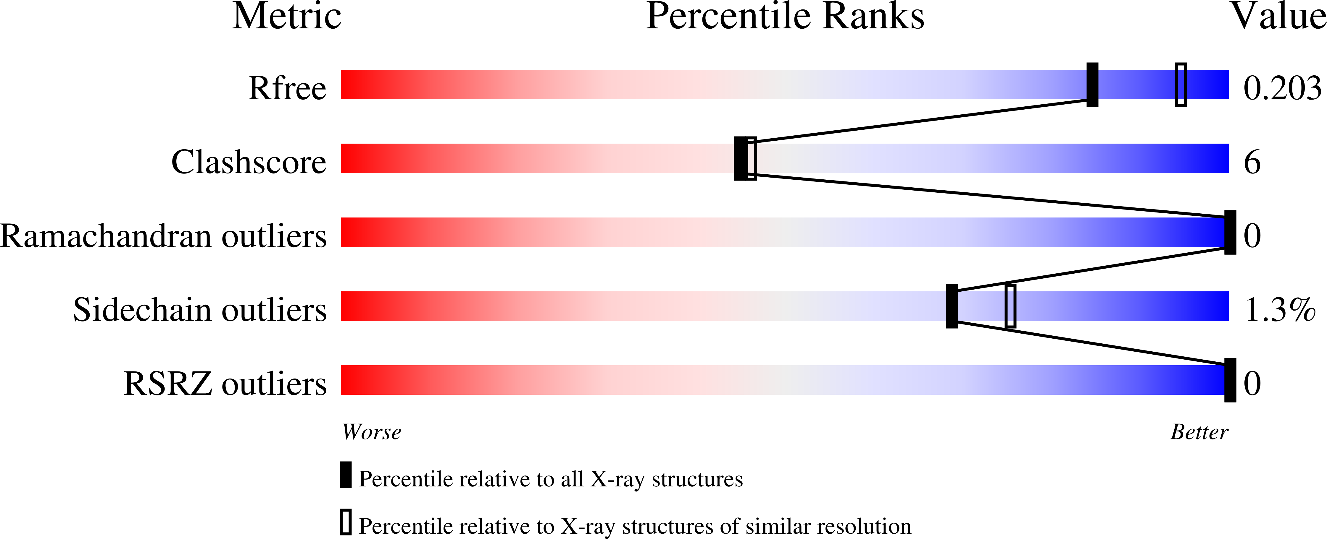
R-Value Free:
0.21
R-Value Work:
0.18
R-Value Observed:
0.18
Space Group:
C 2 2 21