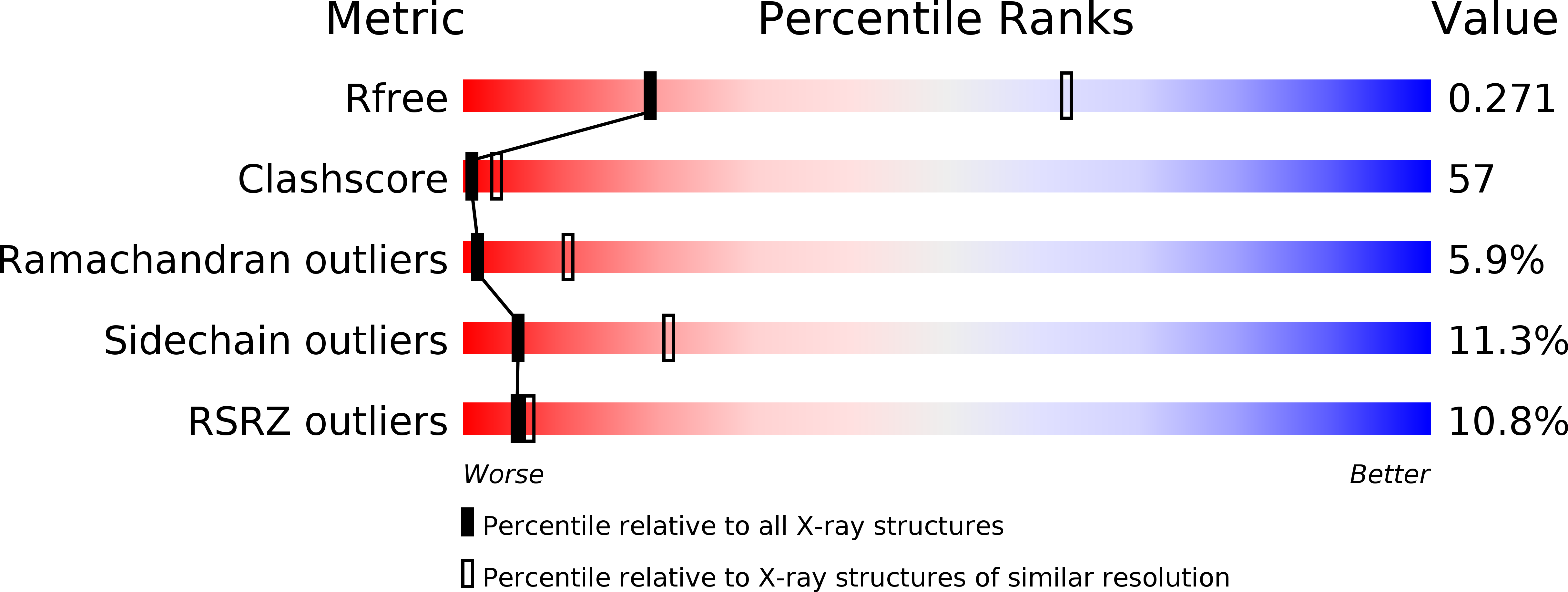
2R4R image
Deposition Date 2007-08-31
Release Date 2007-11-06
Last Version Date 2024-11-06
Entry Detail
PDB ID:
2R4R
Title:
Crystal structure of the human beta2 adrenoceptor
Biological Source:
Source Organism:
Homo sapiens (Taxon ID: 9606)
Mus musculus (Taxon ID: 10090)
Method Details:
Experimental Method:
Resolution:
3.40 Å
R-Value Free:
0.27
R-Value Work:
0.21
Space Group:
C 1 2 1
Macromolecular Entities
Polymer Type:polypeptide(L)
Molecule:Beta-2 adrenergic receptor
Gene (Uniprot):ADRB2
Chain IDs:A
Chain Length:365
Number of Molecules:1
Biological Source:Homo sapiens
Polymer Type:polypeptide(L)
Molecule:antibody for beta2 adrenoceptor, heavy chain
Chain IDs:C (auth: H)
Chain Length:217
Number of Molecules:1
Biological Source:Mus musculus
Polymer Type:polypeptide(L)
Molecule:antibody for beta2 adrenoceptor, light chain
Chain IDs:B (auth: L)
Chain Length:214
Number of Molecules:1
Biological Source:Mus musculus
Ligand Molecules
Primary Citation
Crystal structure of the human beta2 adrenergic G-protein-coupled receptor.
Nature 450 383 387 (2007)
PMID: 17952055 DOI: 10.1038/nature06325

Abstact

Structural analysis of G-protein-coupled receptors (GPCRs) for hormones and neurotransmitters has been hindered by their low natural abundance, inherent structural flexibility, and instability in detergent solutions. Here we report a structure of the human beta2 adrenoceptor (beta2AR), which was crystallized in a lipid environment when bound to an inverse agonist and in complex with a Fab that binds to the third intracellular loop. Diffraction data were obtained by high-brilliance microcrystallography and the structure determined at 3.4 A/3.7 A resolution. The cytoplasmic ends of the beta2AR transmembrane segments and the connecting loops are well resolved, whereas the extracellular regions of the beta2AR are not seen. The beta2AR structure differs from rhodopsin in having weaker interactions between the cytoplasmic ends of transmembrane (TM)3 and TM6, involving the conserved E/DRY sequences. These differences may be responsible for the relatively high basal activity and structural instability of the beta2AR, and contribute to the challenges in obtaining diffraction-quality crystals of non-rhodopsin GPCRs.

Legend

Protein

Chemical

Disease

Primary Citation of related structures