2R4E image
Deposition Date 2007-08-31
Release Date 2008-04-29
Last Version Date 2024-02-21
Entry Detail
PDB ID:
2R4E
Keywords:
Title:
Crystal structure of Escherichia coli Glycerol-3-phosphate Dehydrogenase in complex with DHAP
Biological Source:
Source Organism:
Escherichia coli (Taxon ID: 562)
Host Organism:
Method Details:
Experimental Method:
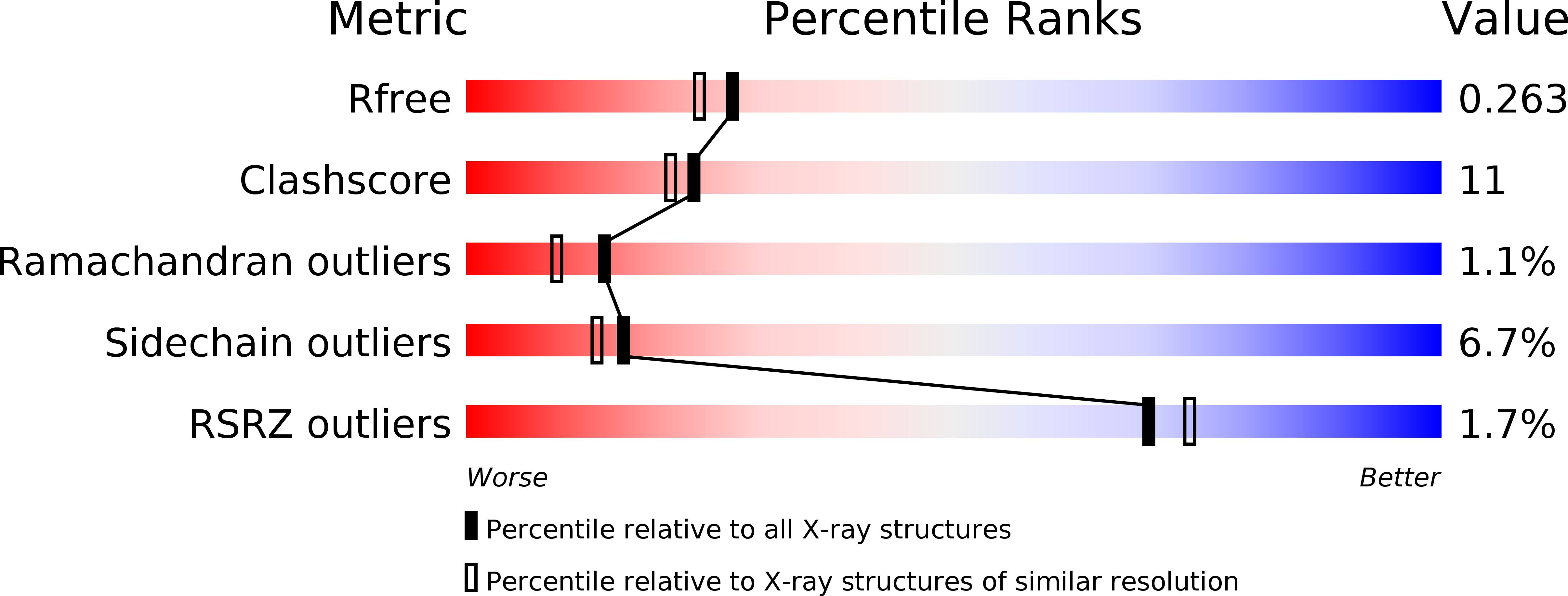
Resolution:
2.10 Å
R-Value Free:
0.25
R-Value Work:
0.19
R-Value Observed:
0.20
Space Group:
I 2 2 2
Macromolecular Entities
Polymer Type:polypeptide(L)
Molecule:Aerobic glycerol-3-phosphate dehydrogenase
Gene (Uniprot):glpD
Chain IDs:A, B
Chain Length:501
Number of Molecules:2
Biological Source:Escherichia coli
Primary Citation
Structure of glycerol-3-phosphate dehydrogenase, an essential monotopic membrane enzyme involved in respiration and metabolism.
Proc.Natl.Acad.Sci.Usa 105 3280 3285 (2008)
PMID: 18296637 DOI: 10.1073/pnas.0712331105

Abstact

Sn-glycerol-3-phosphate dehydrogenase (GlpD) is an essential membrane enzyme, functioning at the central junction of respiration, glycolysis, and phospholipid biosynthesis. Its critical role is indicated by the multitiered regulatory mechanisms that stringently controls its expression and function. Once expressed, GlpD activity is regulated through lipid-enzyme interactions in Escherichia coli. Here, we report seven previously undescribed structures of the fully active E. coli GlpD, up to 1.75 A resolution. In addition to elucidating the structure of the native enzyme, we have determined the structures of GlpD complexed with substrate analogues phosphoenolpyruvate, glyceric acid 2-phosphate, glyceraldehyde-3-phosphate, and product, dihydroxyacetone phosphate. These structural results reveal conformational states of the enzyme, delineating the residues involved in substrate binding and catalysis at the glycerol-3-phosphate site. Two probable mechanisms for catalyzing the dehydrogenation of glycerol-3-phosphate are envisioned, based on the conformational states of the complexes. To further correlate catalytic dehydrogenation to respiration, we have additionally determined the structures of GlpD bound with ubiquinone analogues menadione and 2-n-heptyl-4-hydroxyquinoline N-oxide, identifying a hydrophobic plateau that is likely the ubiquinone-binding site. These structures illuminate probable mechanisms of catalysis and suggest how GlpD shuttles electrons into the respiratory pathway. Glycerol metabolism has been implicated in insulin signaling and perturbations in glycerol uptake and catabolism are linked to obesity in humans. Homologs of GlpD are found in practically all organisms, from prokaryotes to humans, with >45% consensus protein sequences, signifying that these structural results on the prokaryotic enzyme may be readily applied to the eukaryotic GlpD enzymes.

Legend

Protein

Chemical

Disease

Primary Citation of related structures