Deposition Date
2007-08-30
Release Date
2008-09-02
Last Version Date
2025-08-06
Entry Detail
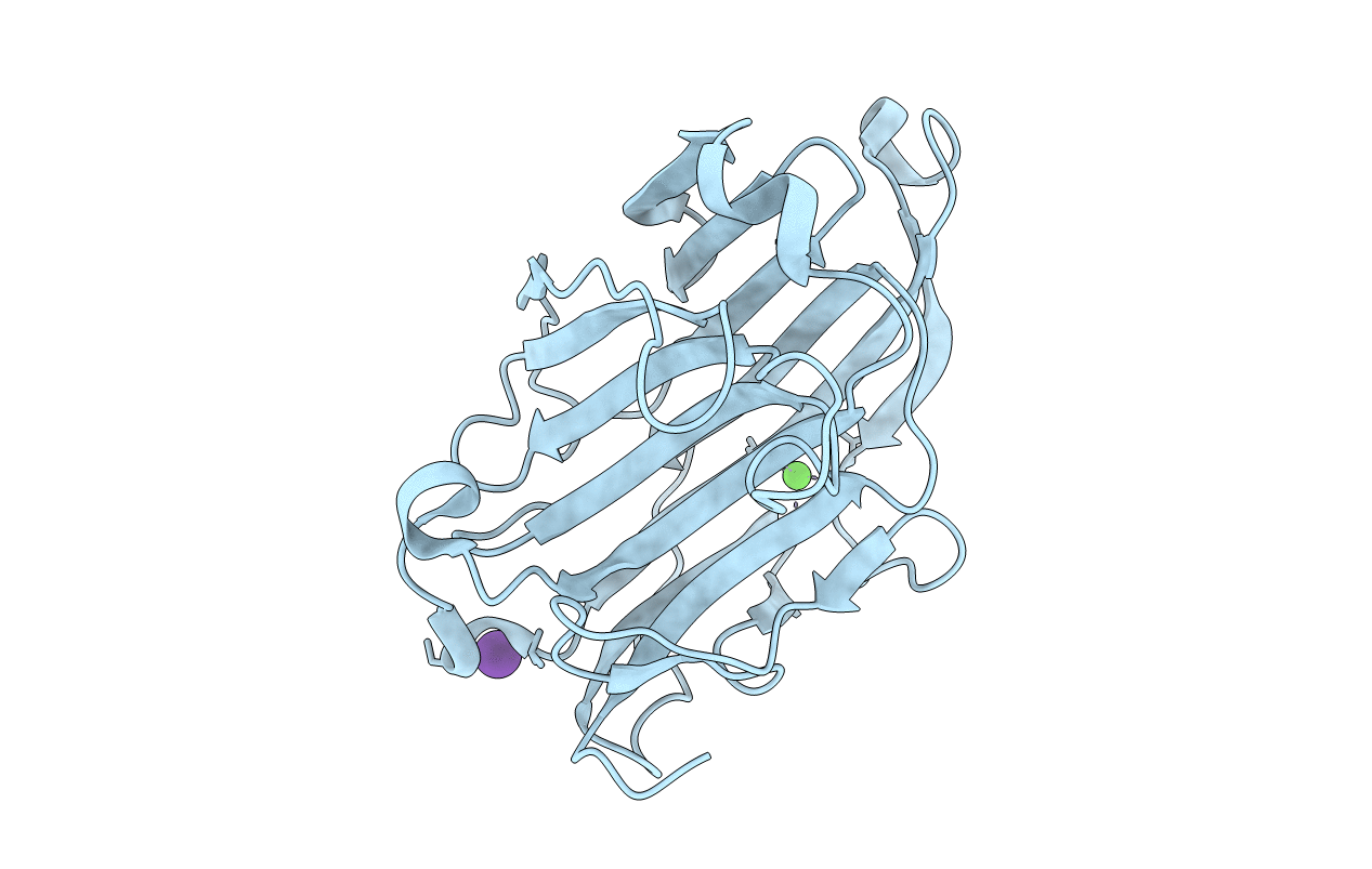
PDB ID:
2R49
Keywords:
Title:
Mutational and Structural Studies of E85I Reveal the Flexible Loops of Fibrobacter succinogenes 1,3-1,4-beta-D-GlucanaseGlucanase
Biological Source:
Source Organism(s):
Fibrobacter succinogenes (Taxon ID: 833)
Expression System(s):
Method Details:
Experimental Method:
Resolution:
2.20 Å
R-Value Free:
0.26
R-Value Work:
0.19
R-Value Observed:
0.19
Space Group:
P 31 2 1