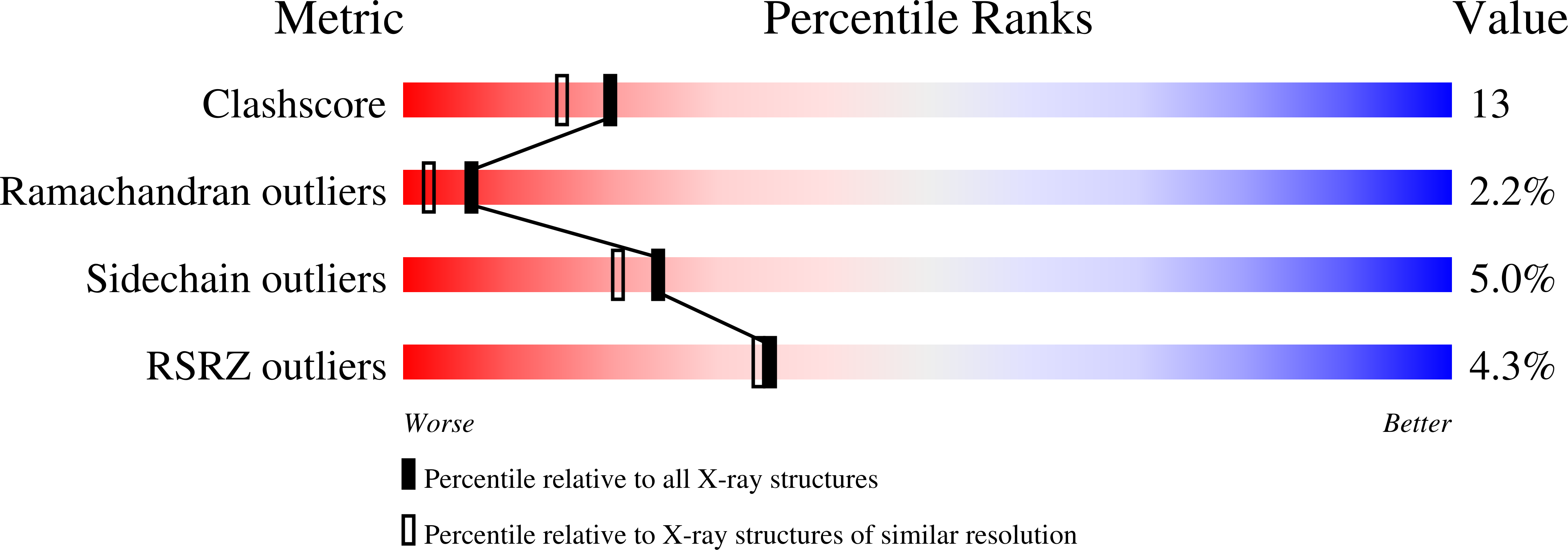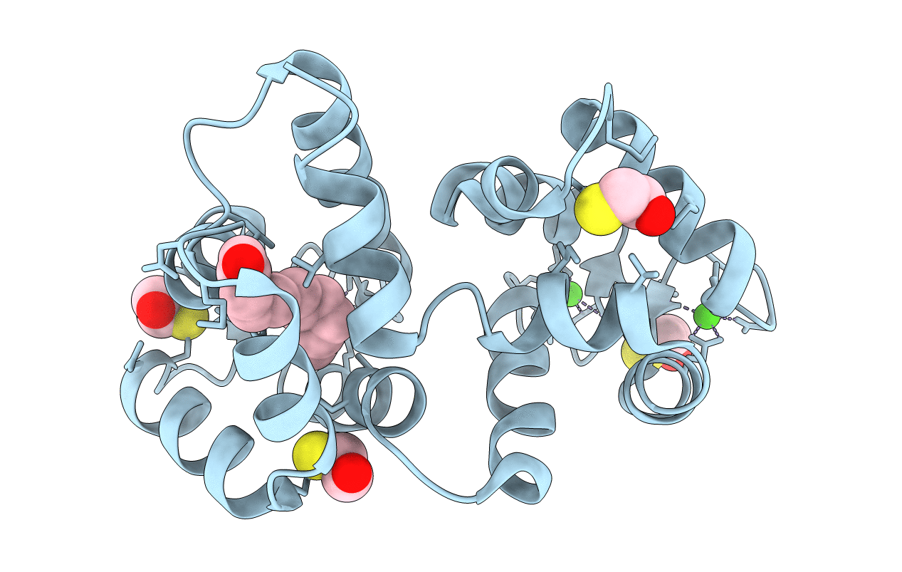
2R2I image
Deposition Date 2007-08-25
Release Date 2007-12-11
Last Version Date 2021-10-20
Entry Detail
PDB ID:
2R2I
Keywords:
Title:
Myristoylated Guanylate Cyclase Activating Protein-1 with Calcium Bound
Biological Source:
Source Organism:
Gallus gallus (Taxon ID: 9031)
Host Organism:
Method Details:
Experimental Method:
Resolution:
2.00 Å
R-Value Free:
0.25
R-Value Work:
0.21
R-Value Observed:
0.21
Space Group:
P 41 21 2
Macromolecular Entities
Polymer Type:polypeptide(L)
Molecule:Guanylyl cyclase-activating protein 1
Gene (Uniprot):GUCA1A
Mutations:G6S
Chain IDs:A
Chain Length:198
Number of Molecules:1
Biological Source:Gallus gallus
Primary Citation
Stabilizing function for myristoyl group revealed by the crystal structure of a neuronal calcium sensor, guanylate cyclase-activating protein 1.
Structure 15 1392 1402 (2007)
PMID: 17997965 DOI: 10.1016/j.str.2007.09.013

Abstact

Guanylate cyclase-activating proteins (GCAPs) are Ca(2+)-binding proteins myristoylated at the N terminus that regulate guanylate cyclases in photoreceptor cells and belong to the family of neuronal calcium sensors (NCS). Many NCS proteins display a recoverin-like "calcium-myristoyl switch" whereby the myristoyl group, buried inside the protein in the Ca(2+)-free state, becomes fully exposed upon Ca(2+) binding. Here we present a 2.0 A resolution crystal structure of myristoylated GCAP1 with Ca(2+) bound. The acyl group is buried inside Ca(2+)-bound GCAP1. This is in sharp contrast to Ca(2+)-bound recoverin, where the myristoyl group is solvent exposed. Furthermore, we provide direct evidence that the acyl group in GCAP1 remains buried in the Ca(2+)-free state and does not undergo switching. A pronounced kink in the C-terminal helix and the presence of the myristoyl group allow clustering of sequence elements crucial for GCAP1 activity.

Legend

Protein

Chemical

Disease

Primary Citation of related structures