2R28 image
Deposition Date 2007-08-24
Release Date 2008-07-01
Last Version Date 2023-08-30
Entry Detail
PDB ID:
2R28
Title:
The complex Structure of Calmodulin Bound to a Calcineurin Peptide
Biological Source:
Source Organism:
Homo sapiens (Taxon ID: )
Host Organism:
Method Details:
Experimental Method:
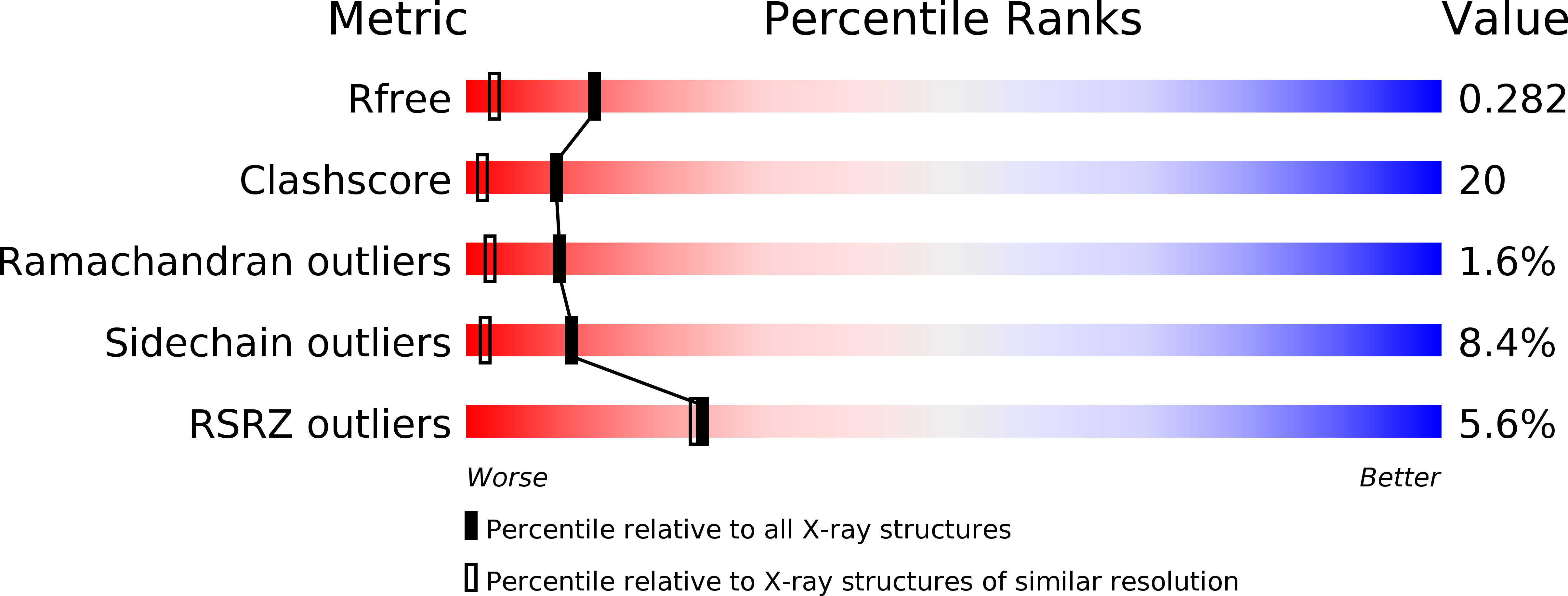
Resolution:
1.86 Å
R-Value Free:
0.28
R-Value Work:
0.23
R-Value Observed:
0.23
Space Group:
C 1 2 1
Macromolecular Entities
Structures with similar UniProt ID
Protein Blast
Polymer Type:polypeptide(L)
Molecule:Calmodulin
Chain IDs:A, C (auth: B)
Chain Length:149
Number of Molecules:2
Biological Source:Homo sapiens
Structures with similar UniProt ID
Protein Blast
Polymer Type:polypeptide(L)
Molecule:Serine/threonine-protein phosphatase 2B catalytic subunit alpha isoform
Gene (Uniprot):PPP3CA
Chain IDs:B (auth: C), D
Chain Length:25
Number of Molecules:2
Biological Source:Homo sapiens
Ligand Molecules
Primary Citation
The complex structure of calmodulin bound to a calcineurin peptide.
Proteins 73 19 27 (2008)
PMID: 18384083 DOI: 10.1002/prot.22032

Abstact

The activity of the protein phosphatase calcineurin (CN) is regulated by an autoinhibition mechanism wherein several domains from its catalytic A subunit, including the calmodulin binding domain (CaMBD), block access to its active site. Upon binding of Ca2+ and calmodulin (Ca2+/CaM) to CaMBD, the autoinhibitory domains dissociate from the catalytic groove, thus activating the enzyme. To date, the structure of the CN/CaM/Ca2+ complex has not been determined in its entirety. Previously, we determined the structure of a fusion protein consisting of CaM and a 25-residue peptide taken from the CaMBD, joined by a 5-glycine linker. This structure revealed a novel CaM binding motif. However, the presence of the extraneous glycine linker cast doubt on the authenticity of this structure as an accurate representation of CN/CaM binding in vivo. Thus, here, we have determined the crystal structure of CaM complexed with the 25-residue CaMBD peptide without the glycine linker at a resolution of 2.1 A. The structure is essentially identical to the fusion construction which displays CaM bound to the CaMBD peptide as a dimer with an open, elongated conformation. The N-lobe from one molecule and C-lobe from another encompass and bind the CaMBD peptide. Thus, it validates the existence of this novel CaM binding motif. Our experiments suggest that the dimeric CaM/CaMBD complex exists in solution, which is unambiguously validated using a carefully-designed CaM-sepharose pull-down experiment. We discuss structural features that produce this novel binding motif, including the role of the CaMBD peptide residues Arg-408, Val-409, and Phe-410, which work to provide rigidity to the otherwise flexible central CaM helix joining the N- and C-lobes, ultimately keeping these lobes apart and forcing "head-to-tail" dimerization to attain the requisite N- and C-lobe pairing for CaMBD binding.

Legend

Protein

Chemical

Disease

Primary Citation of related structures