2R25 image
Entry Detail
PDB ID:
2R25
Title:
Complex of YPD1 and SLN1-R1 with bound Mg2+ and BeF3-
Biological Source:
Source Organism:
Host Organism:
PDB Version:
Deposition Date:
2007-08-24
Release Date:
2008-01-15
Method Details:
Experimental Method:
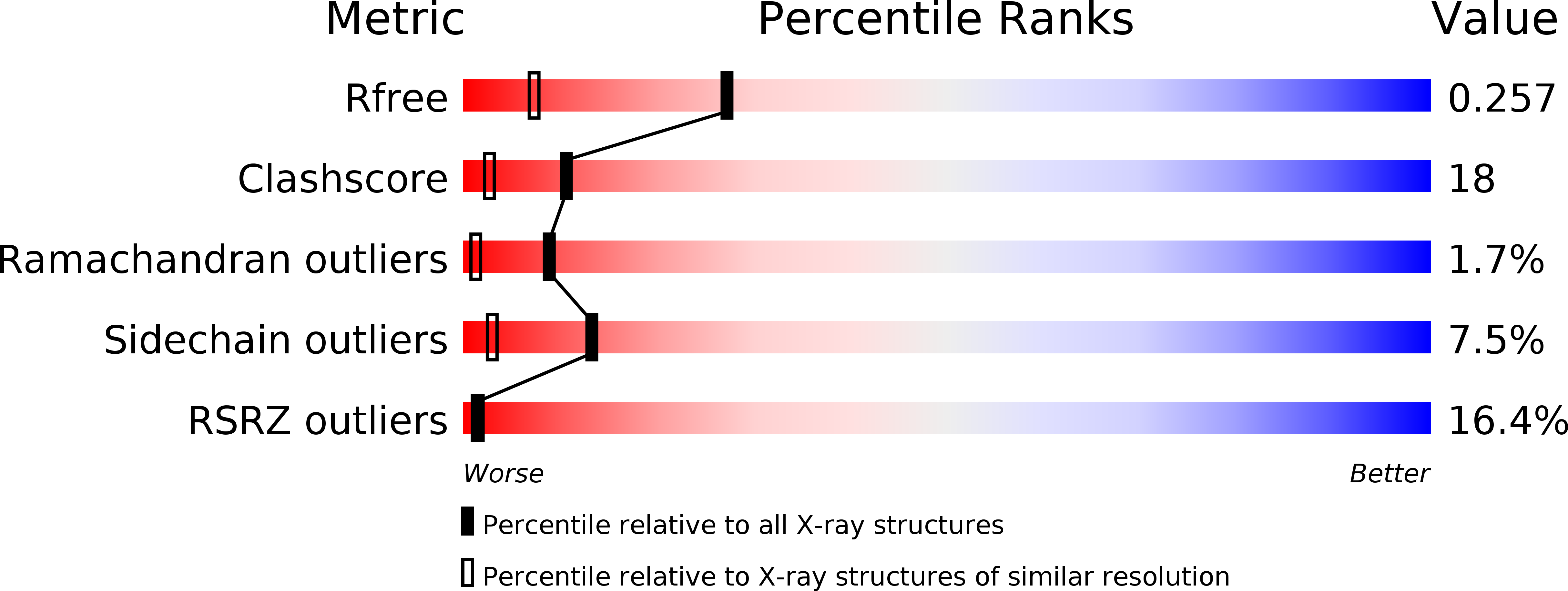
Resolution:
1.70 Å
R-Value Free:
0.24
R-Value Work:
0.19
R-Value Observed:
0.19
Space Group:
P 21 21 21
Macromolecular Entities
Polymer Type:polypeptide(L)
Description:Phosphorelay intermediate protein YPD1
Chain IDs:A
Chain Length:167
Number of Molecules:1
Biological Source:Saccharomyces cerevisiae
Polymer Type:polypeptide(L)
Description:Osmosensing histidine protein kinase SLN1
Chain IDs:B
Chain Length:133
Number of Molecules:1
Biological Source:Saccharomyces cerevisiae
Primary Citation
Crystal structure of a complex between the phosphorelay protein YPD1 and the response regulator domain of SLN1 bound to a phosphoryl analog
J.Mol.Biol. 375 1141 1151 (2008)
PMID: 18076904 DOI: 10.1016/j.jmb.2007.11.045

Abstact

The crystal structure of the yeast SLN1 response regulator (RR) domain bound to both a phosphoryl analog [beryllium fluoride (BeF(3)(-))] and Mg(2+), in complex with its downstream phosphorelay signaling partner YPD1, has been determined at a resolution of 1.70 A. Comparisons between the BeF(3)(-)-activated complex and the unliganded (or apo) complex determined previously reveal modest but important differences. The SLN1-R1 x Mg(2+) x BeF(3)(-) structure from the complex provides evidence for the first time that the mechanism of phosphorylation-induced activation is highly conserved between bacterial RR domains and this example from a eukaryotic organism. Residues in and around the active site undergo slight rearrangements in order to form bonds with the essential divalent cation and fluorine atoms of BeF(3)(-). Two conserved switch-like residues (Thr1173 and Phe1192) occupy distinctly different positions in the apo versus BeF(3)(-)-bound structures, consistent with the "Y-T" coupling mechanism proposed for the activation of CheY and other bacterial RRs. Several loop regions and the alpha 4-beta 5-alpha 5 surface of the SLN1-R1 domain undergo subtle conformational changes (approximately 1-3 A displacements relative to the apo structure) that lead to significant changes in terms of contacts that are formed with YPD1. Detailed structural comparisons of protein-protein interactions in the apo and BeF(3)(-)-bound complexes suggest at least a two-state equilibrium model for the formation of a transient encounter complex, in which phosphorylation of the RR promotes the formation of a phosphotransfer-competent complex. In the BeF(3)(-)-activated complex, the position of His64 from YPD1 needs to be within ideal distance of and in near-linear geometry with Asp1144 from the SLN1-R1 domain for phosphotransfer to occur. The ground-state structure presented here suggests that phosphoryl transfer will likely proceed through an associative mechanism involving the formation of a pentacoordinate phosphorus intermediate.

Legend

Protein

Chemical

Disease

Primary Citation of related structures