2R1B image
Deposition Date 2007-08-22
Release Date 2008-03-25
Last Version Date 2023-08-30
Entry Detail
PDB ID:
2R1B
Title:
Crystal Structure of rat neurexin 1beta with a splice insert at SS#4
Biological Source:
Source Organism:
Method Details:
Experimental Method:
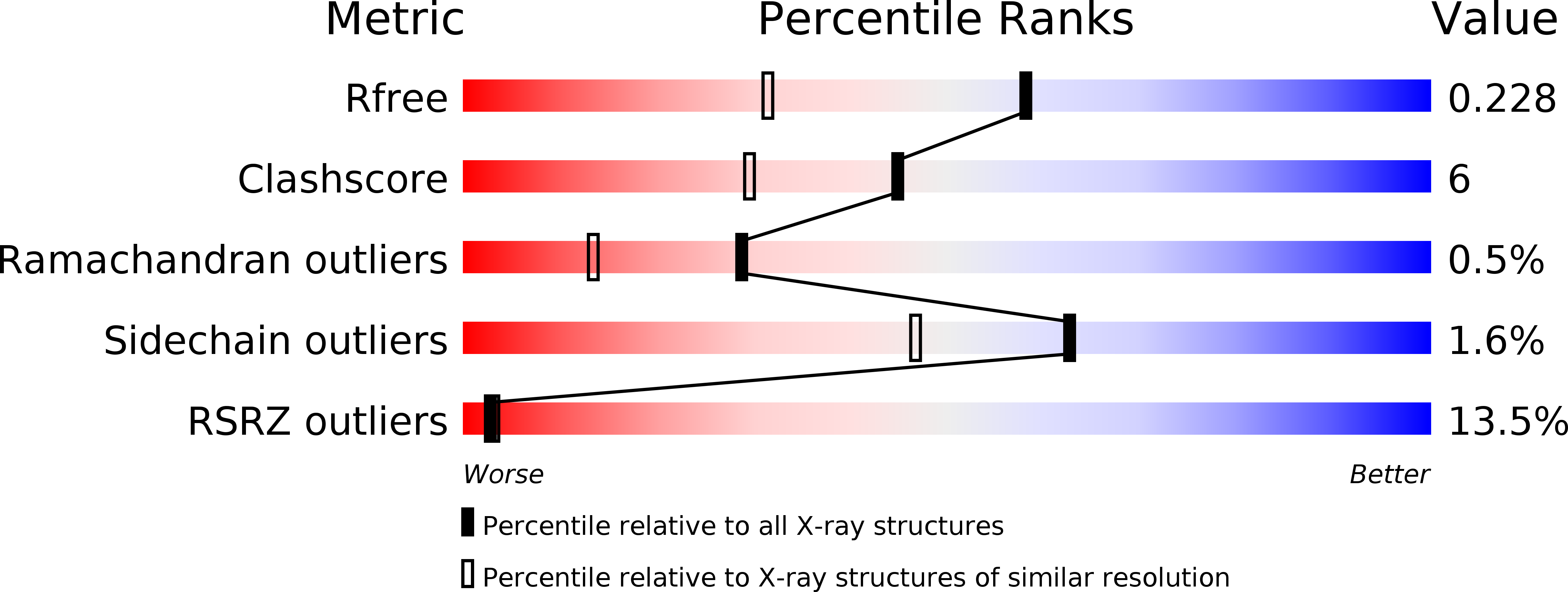
Resolution:
1.72 Å
R-Value Free:
0.22
R-Value Work:
0.19
R-Value Observed:
0.19
Space Group:
P 1 21 1
Macromolecular Entities
Structures with similar UniProt ID
Protein Blast
Polymer Type:polypeptide(L)
Molecule:Neurexin-1-beta
Gene (Uniprot):Nrxn1
Chain IDs:A, B
Chain Length:220
Number of Molecules:2
Biological Source:Rattus norvegicus
Ligand Molecules
Primary Citation
Regulation of Neurexin 1beta Tertiary Structure and Ligand Binding through Alternative Splicing
Structure 16 422 431 (2008)
PMID: 18334217 DOI: 10.1016/j.str.2008.01.005

Abstact

Neurexins and neuroligins play an essential role in synapse function, and their alterations are linked to autistic spectrum disorder. Interactions between neurexins and neuroligins regulate inhibitory and excitatory synaptogenesis in vitro through a "splice-insert signaling code." In particular, neurexin 1beta carrying an alternative splice insert at site SS#4 interacts with neuroligin 2 (found predominantly at inhibitory synapses) but much less so with other neuroligins (those carrying an insert at site B and prevalent at excitatory synapses). The structure of neurexin 1beta+SS#4 reveals dramatic rearrangements to the "hypervariable surface," the binding site for neuroligins. The splice insert protrudes as a long helix into space, triggers conversion of loop beta10-beta11 into a helix rearranging the binding site for neuroligins, and rearranges the Ca(2+)-binding site required for ligand binding, increasing its affinity. Our structures reveal the mechanism by which neurexin 1beta isoforms acquire neuroligin splice isoform selectivity.

Legend

Protein

Chemical

Disease

Primary Citation of related structures
Feedback Form
Name
Email
Institute
Feedback