2R13 image
Deposition Date 2007-08-22
Release Date 2007-09-11
Last Version Date 2024-03-13
Entry Detail
PDB ID:
2R13
Title:
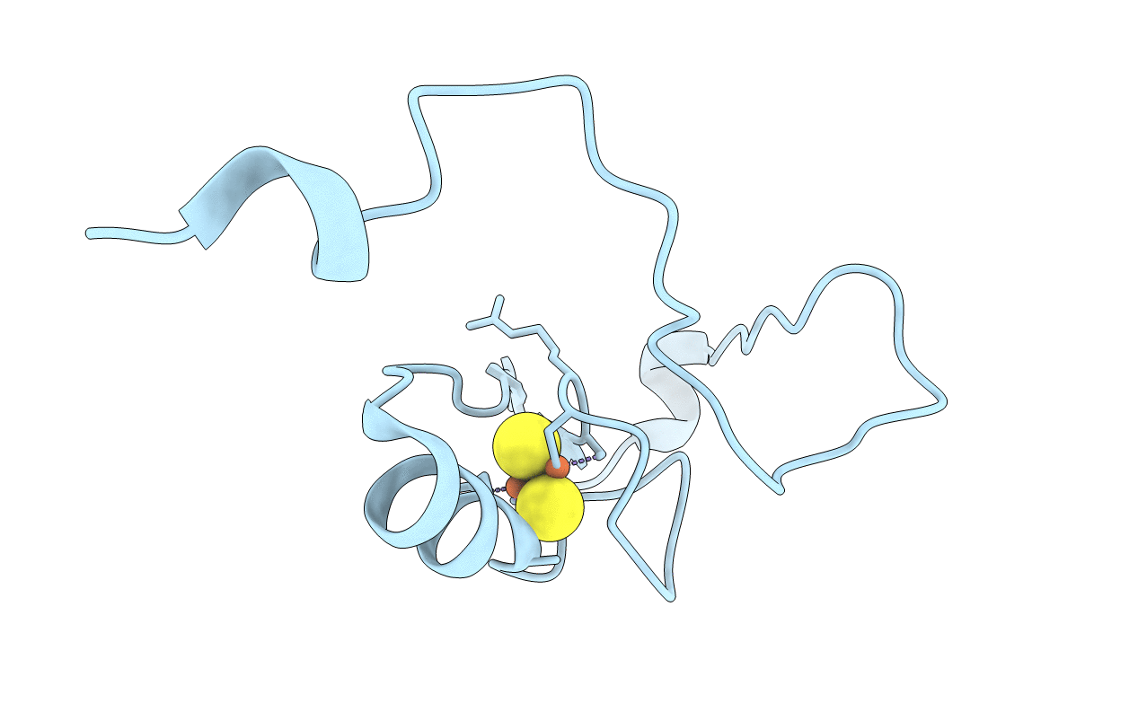
Crystal structure of human mitoNEET reveals a novel [2Fe-2S] cluster coordination
Biological Source:
Source Organism:
Homo sapiens (Taxon ID: 9606)
Host Organism:
Method Details:
Experimental Method:
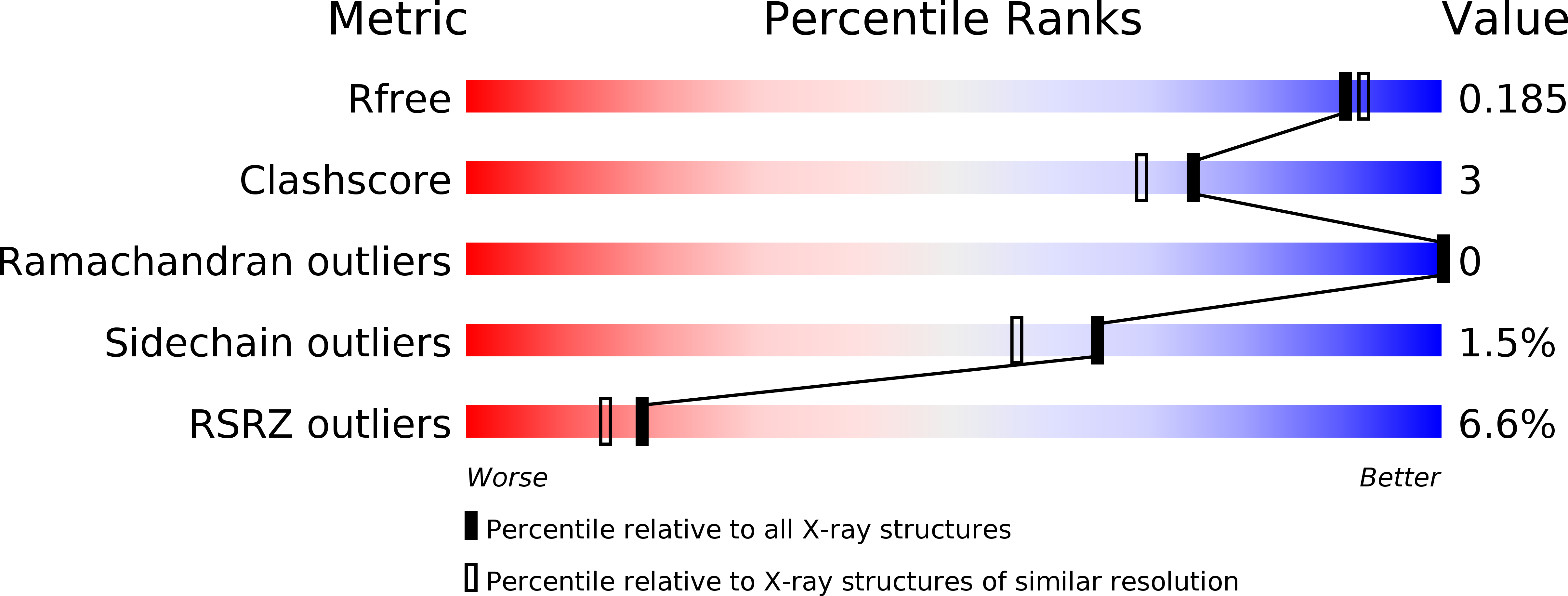
Resolution:
1.80 Å
R-Value Free:
0.18
R-Value Work:
0.15
R-Value Observed:
0.16
Space Group:
I 41 2 2
Macromolecular Entities
Structures with similar UniProt ID
Protein Blast
Polymer Type:polypeptide(L)
Molecule:Zinc finger CDGSH domain-containing protein 1
Gene (Uniprot):CISD1
Chain IDs:A
Chain Length:79
Number of Molecules:1
Biological Source:Homo sapiens
Primary Citation
Crystallographic studies of human MitoNEET
J.Biol.Chem. 282 33242 33246 (2007)
PMID: 17905743 DOI: 10.1074/jbc.C700172200

Abstact

MitoNEET was identified as an outer mitochondrial membrane protein that can potentially bind the anti-diabetes drug pioglitazone. The crystal structure of the cytoplasmic mitoNEET (residues 33-108) is determined in this study. The structure presents a novel protein fold and contains a [2Fe-2S] cluster-binding domain. The [2Fe-2S] cluster is coordinated to the protein by Cys-72, Cys-74, Cys-83, and His-87 residues. This coordination is also novel compared with the traditional [2Fe-2S] cluster coordinated by four cysteines or two cysteines and two histidines. The cytoplasmic mitoNEET forms homodimers in solution and in crystal. The dimerization is mainly mediated by hydrophobic interactions as well as hydrogen bonds coordinated by two water molecules binding at the interface. His-87 residue, which plays an important role in the coordination of the [2Fe-2S] cluster, is exposed to the solvent on the dimer surface. It is proposed that mitoNEET dimer may interact with other proteins via the surface residues in close proximity to the [2Fe-2S] cluster.

Legend

Protein

Chemical

Disease

Primary Citation of related structures