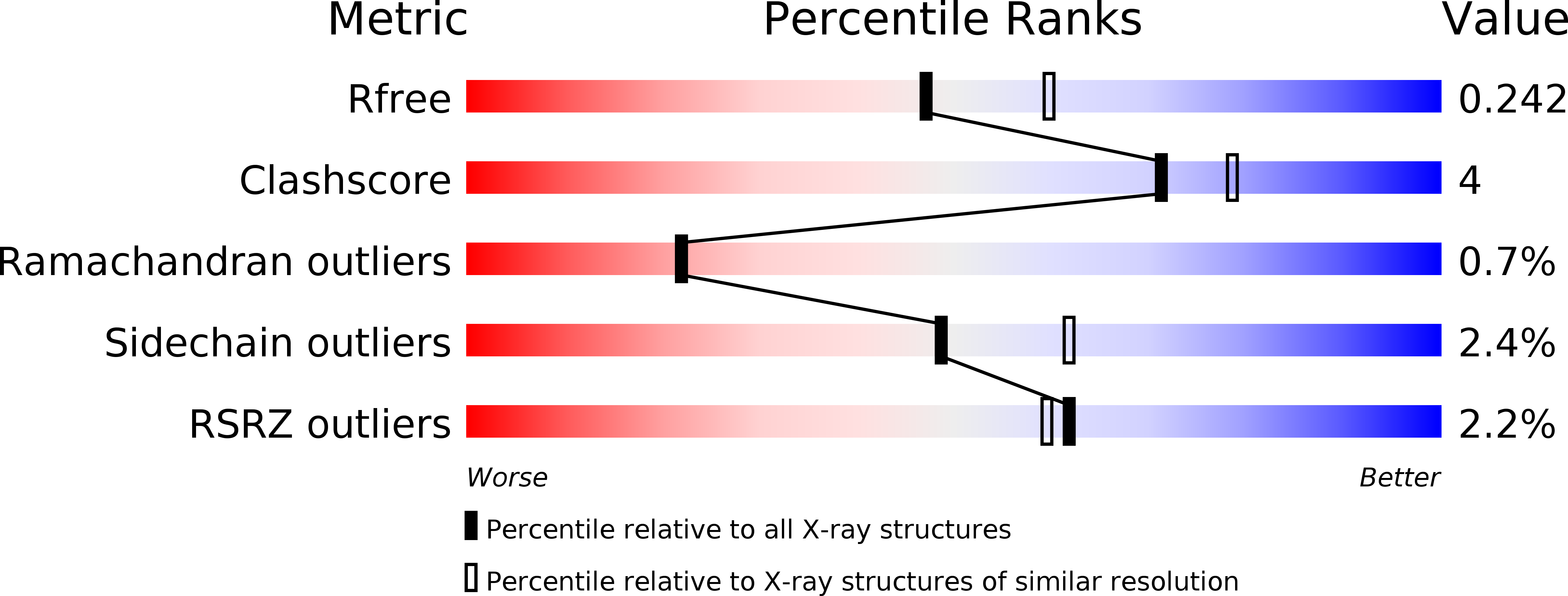
2R0L image
Deposition Date 2007-08-20
Release Date 2007-12-25
Last Version Date 2024-11-06
Entry Detail
PDB ID:
2R0L
Title:
Short Form HGFA with Inhibitory Fab75
Biological Source:
Source Organism:
Host Organism:
Method Details:
Experimental Method:
Resolution:
2.20 Å
R-Value Free:
0.24
R-Value Work:
0.19
R-Value Observed:
0.19
Space Group:
P 1
Macromolecular Entities
Polymer Type:polypeptide(L)
Molecule:Hepatocyte growth factor activator
Gene (Uniprot):HGFAC
Chain IDs:C (auth: A)
Chain Length:248
Number of Molecules:1
Biological Source:Homo sapiens
Polymer Type:polypeptide(L)
Molecule:Hepatocyte growth factor activator
Gene (Uniprot):HGFAC
Chain IDs:D (auth: B)
Chain Length:35
Number of Molecules:1
Biological Source:Homo sapiens
Polymer Type:polypeptide(L)
Molecule:antibody heavy chain, Fab portion only
Chain IDs:B (auth: H)
Chain Length:220
Number of Molecules:1
Biological Source:Homo sapiens, Synthetic construct
Polymer Type:polypeptide(L)
Molecule:antibody light chain
Chain IDs:A (auth: L)
Chain Length:214
Number of Molecules:1
Biological Source:Homo sapiens, Synthetic construct
Primary Citation
Structural insight into distinct mechanisms of protease inhibition by antibodies.
Proc.Natl.Acad.Sci.Usa 104 19784 19789 (2007)
PMID: 18077410 DOI: 10.1073/pnas.0708251104

Abstact

To better understand how the relatively flat antigen-combining sites of antibodies interact with the concave shaped substrate-binding clefts of proteases, we determined the structures of two antibodies in complex with the trypsin-like hepatocyte growth-factor activator (HGFA). The two inhibitory antibodies, Ab58 and Ab75, were generated from a human Fab phage display library with synthetic diversity in the three complementarity determining regions (H1, H2, and H3) of the heavy chain, mimicking the natural diversity of the human Ig repertoire. Biochemical studies and the structures of the Fab58:HGFA (3.5-A resolution) and the Fab75:HGFA (2.2-A resolution) complexes revealed that Ab58 obstructed substrate access to the active site, whereas Ab75 allosterically inhibited substrate hydrolysis. In both cases, the antibodies interacted with the same protruding element (99-loop), which forms part of the substrate-binding cleft. Ab58 inserted its H1 and H2 loops in the cleft to occupy important substrate interaction sites (S3 and S2). In contrast, Ab75 bound at the backside of the cleft to a region corresponding to thrombin exosite II, which is known to interact with allosteric effector molecules. In agreement with the structural analysis, binding assays with active site inhibitors and enzymatic assays showed that Ab58 is a competitive inhibitor, and Ab75 is a partial competitive inhibitor. These results provide structural insight into antibody-mediated protease inhibition. They suggest that unlike canonical inhibitors, antibodies may preferentially target protruding loops at the rim of the substrate-binding cleft to interfere with the catalytic machinery of proteases without requiring long insertion loops.

Legend

Protein

Chemical

Disease

Primary Citation of related structures