2QXG image
Entry Detail
PDB ID:
2QXG
Keywords:
Title:
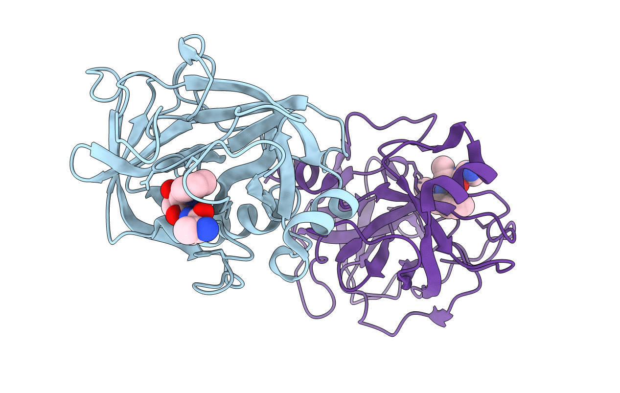
Crystal Structure of Human Kallikrein 7 in Complex with Ala-Ala-Phe-chloromethylketone
Biological Source:
Source Organism:
Host Organism:
PDB Version:
Deposition Date:
2007-08-11
Release Date:
2008-01-08
Method Details:
Experimental Method:
Resolution:
2.60 Å
R-Value Free:
0.29
R-Value Work:
0.26
R-Value Observed:
0.26
Space Group:
P 1 21 1
Macromolecular Entities
Polymer Type:polypeptide(L)
Description:Kallikrein-7
Chain IDs:A, B
Chain Length:224
Number of Molecules:2
Biological Source:Homo sapiens
Ligand Molecules
Primary Citation
Chymotryptic specificity determinants in the 1.0 A structure of the zinc-inhibited human tissue kallikrein 7.
Proc.Natl.Acad.Sci.Usa 104 16086 16091 (2007)
PMID: 17909180 DOI: 10.1073/pnas.0707811104

Abstact

hK7 or human stratum corneum chymotryptic enzyme belongs to the human tissue kallikrein (hKs) serine proteinase family and is strongly expressed in the upper layers of the epidermis. It participates in skin desquamation but is also implicated in diverse skin diseases and is a potential biomarker of ovarian cancer. We have solved x-ray structures of recombinant active hK7 at medium and atomic resolution in the presence of the inhibitors succinyl-Ala-Ala-Pro-Phe-chloromethyl ketone and Ala-Ala-Phe-chloromethyl ketone. The most distinguishing features of hK7 are the short 70-80 loop and the unique S1 pocket, which prefers P1 Tyr residues, as shown by kinetic data. Similar to several other kallikreins, the enzyme activity is inhibited by Zn(2+) and Cu(2+) at low micromolar concentrations. Biochemical analyses of the mutants H99A and H41F confirm that only the metal-binding site at His(99) close to the catalytic triad accounts for the noncompetitive Zn(2+) inhibition type. Additionally, hK7 exhibits large positively charged surface patches, representing putative exosites for prime side substrate recognition.

Legend

Protein

Chemical

Disease

Primary Citation of related structures