Deposition Date
2007-08-10
Release Date
2008-03-18
Last Version Date
2023-08-30
Entry Detail
PDB ID:
2QWS
Keywords:
Title:
Neutron and X-ray structural studies of short hydrogen bonds in Photoactive Yellow Protein (PYP)
Biological Source:
Source Organism(s):
Halorhodospira halophila (Taxon ID: 1053)
Method Details:
Experimental Method:
Resolution:
2.50 Å
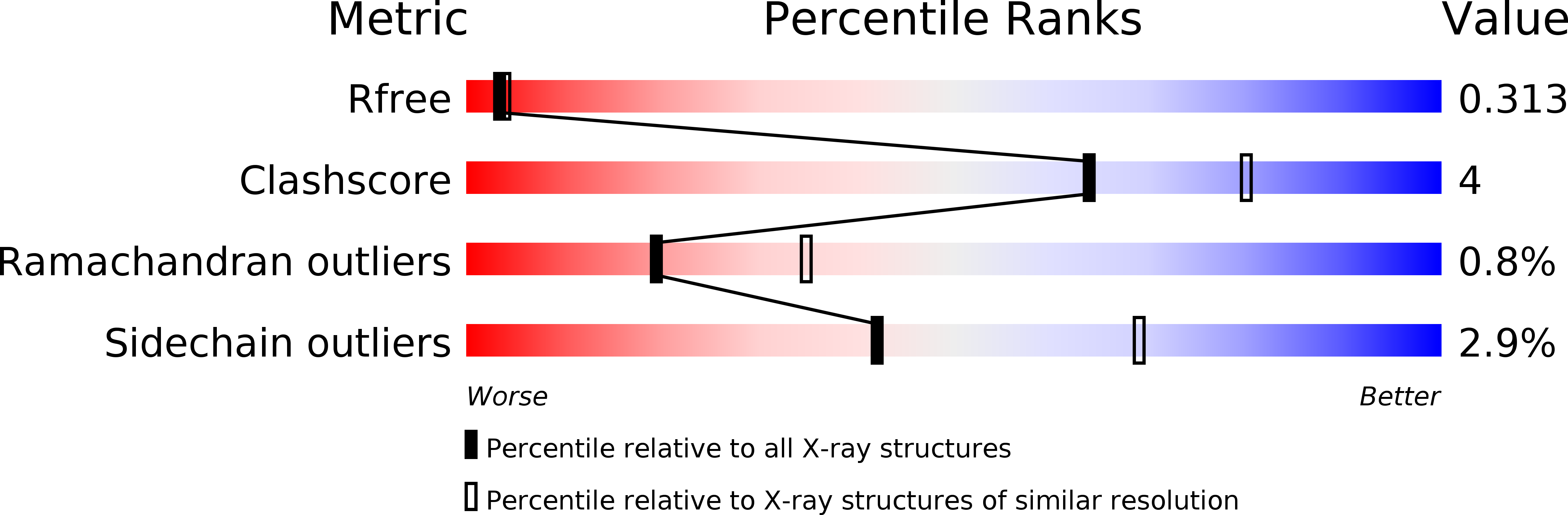
R-Value Free:
0.27
R-Value Work:
0.26
Space Group:
P 63