2QVL image
Deposition Date 2007-08-08
Release Date 2008-06-17
Last Version Date 2024-10-30
Entry Detail
PDB ID:
2QVL
Keywords:
Title:
Crystal Structure of Diacylglycerol Kinase
Biological Source:
Source Organism:
Host Organism:
Method Details:
Experimental Method:
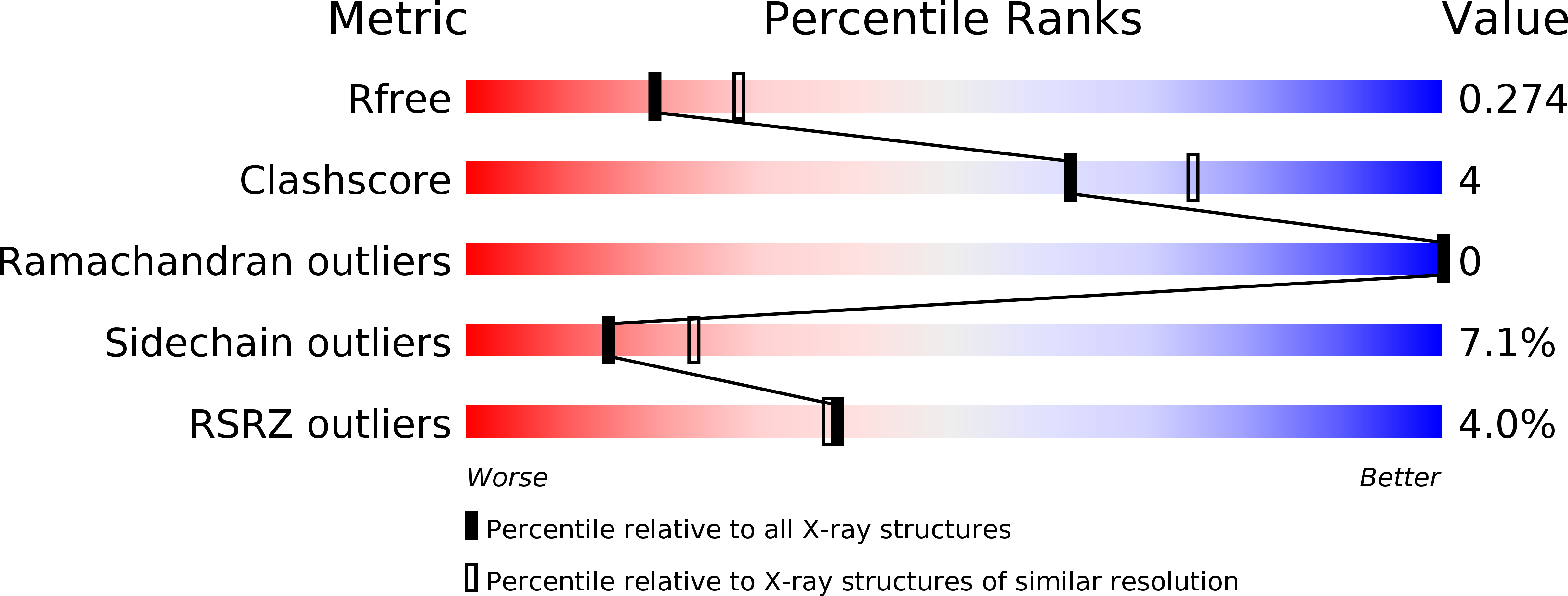
Resolution:
2.40 Å
R-Value Free:
0.27
R-Value Work:
0.22
R-Value Observed:
0.23
Space Group:
P 42 21 2
Macromolecular Entities
Polymer Type:polypeptide(L)
Molecule:Diacylglycerol Kinase DgkB
Gene (Uniprot):dagK
Chain IDs:A
Chain Length:337
Number of Molecules:1
Biological Source:Staphylococcus aureus
Modified Residue
Compound ID Chain ID Parent Comp ID Details 2D Image
MSE A MET SELENOMETHIONINE
Primary Citation
Analysis of the Staphylococcus aureus DgkB Structure Reveals a Common Catalytic Mechanism for the Soluble Diacylglycerol Kinases.
Structure 16 1036 1046 (2008)
PMID: 18611377 DOI: 10.1016/j.str.2008.03.019

Abstact

Soluble diacylglycerol (DAG) kinases function as regulators of diacylglycerol metabolism in cell signaling and intermediary metabolism. We report the structure of a DAG kinase, DgkB from Staphylococcus aureus, both as the free enzyme and in complex with ADP. The molecule is a tight homodimer, and each monomer comprises two domains with the catalytic center located within the interdomain cleft. Two distinctive features of DkgB are a structural Mg2+ site and an associated Asp*water*Mg2+ network that extends toward the active site locale. Site-directed mutagenesis revealed that these features play important roles in the catalytic mechanism. The key active site residues and the components of the Asp*water*Mg2+ network are conserved in the catalytic cores of the mammalian signaling DAG kinases, indicating that these enzymes use the same mechanism and have similar structures as DgkB.

Legend

Protein

Chemical

Disease

Primary Citation of related structures