2QUQ image
Deposition Date 2007-08-06
Release Date 2007-11-13
Last Version Date 2024-02-21
Entry Detail
PDB ID:
2QUQ
Title:
Crystal Structure of the Essential Inner Kinetochore Protein Cep3p
Biological Source:
Source Organism:
Host Organism:
Method Details:
Experimental Method:
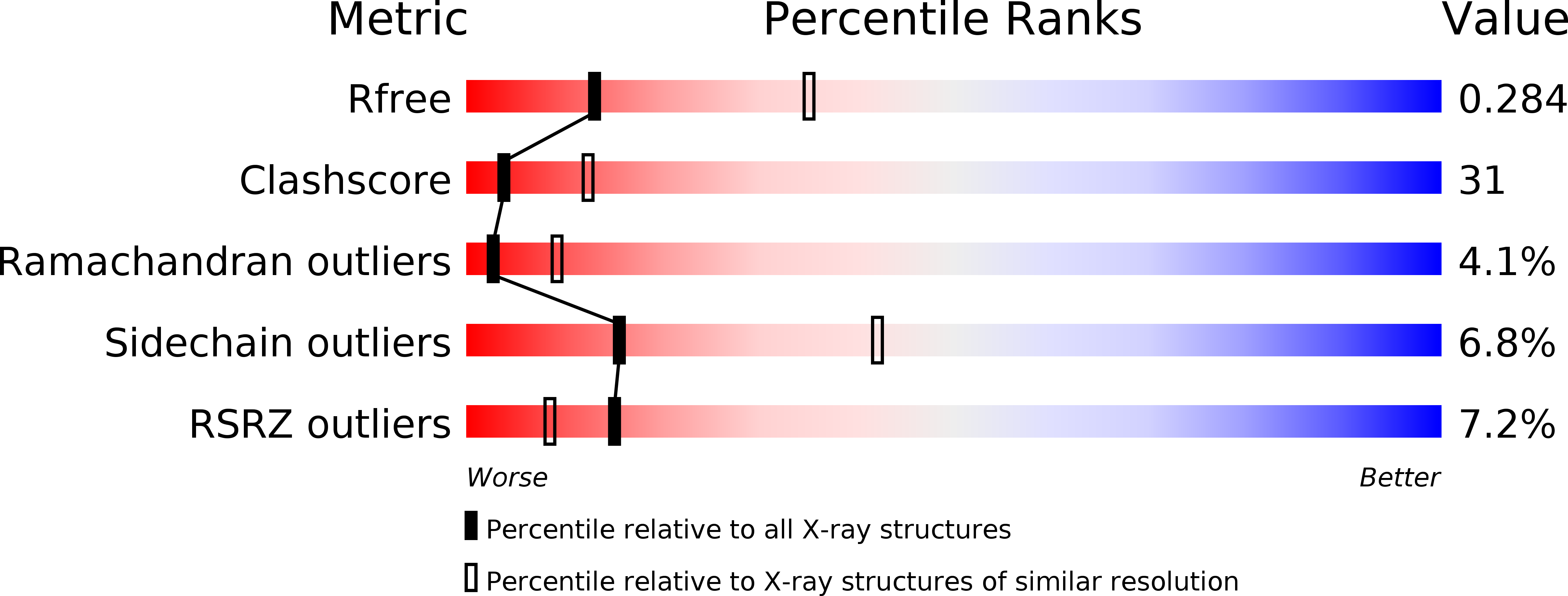
Resolution:
2.80 Å
R-Value Free:
0.28
R-Value Work:
0.22
Space Group:
P 43 21 2
Macromolecular Entities
Structures with similar UniProt ID
Protein Blast
Polymer Type:polypeptide(L)
Molecule:Centromere DNA-binding protein complex CBF3 subunit B
Gene (Uniprot):CEP3
Chain IDs:A
Chain Length:562
Number of Molecules:1
Biological Source:Saccharomyces cerevisiae
Primary Citation
Crystal structure of the yeast inner kinetochore subunit Cep3p.
Structure 15 1422 1430 (2007)
PMID: 17997968 DOI: 10.1016/j.str.2007.09.008

Abstact

In budding yeast, the four-protein CBF3 complex (Skp1p-Ctf13p-Cep3p-Ndc10p) initiates kinetochore assembly by binding to the CDEIII locus of centromeric DNA. A Cep3p dimer recruits a Skp1p-Ctf13p heterodimer and contacts two sites on CDEIII. We report here the crystal structure, determined at 2.8 A resolution by multiple isomorphous replacement with anomalous scattering, of a truncated Cep3p (Cep3p [47-608]), comprising all but an N-terminal, Zn(2)Cys(6)-cluster, DNA-binding module. Cep3p has a well-ordered structure throughout essentially all of its polypeptide chain, unlike most yeast transcription factors, including those with Zn(2)Cys(6) clusters, such as Gal4p. This difference may reflect an underlying functional distinction: whereas any particular transcription factor must adapt to a variety of upstream activating sites, Cep3p scaffolds kinetochore assembly on centromeres uniformly configured on all 16 yeast chromosomes. We have, using the structure of Cep3p (47-608) and the known structures of Zn(2)Cys(6)-cluster domains, modeled the interaction of Cep3p with CDEIII.

Legend

Protein

Chemical

Disease

Primary Citation of related structures