Deposition Date
2007-08-04
Release Date
2007-08-28
Last Version Date
2024-02-21
Entry Detail
PDB ID:
2QUB
Keywords:
Title:
Crystal structure of extracellular lipase LipA from Serratia marcescens
Biological Source:
Source Organism(s):
Serratia marcescens (Taxon ID: 615)
Expression System(s):
Method Details:
Experimental Method:
Resolution:
1.80 Å
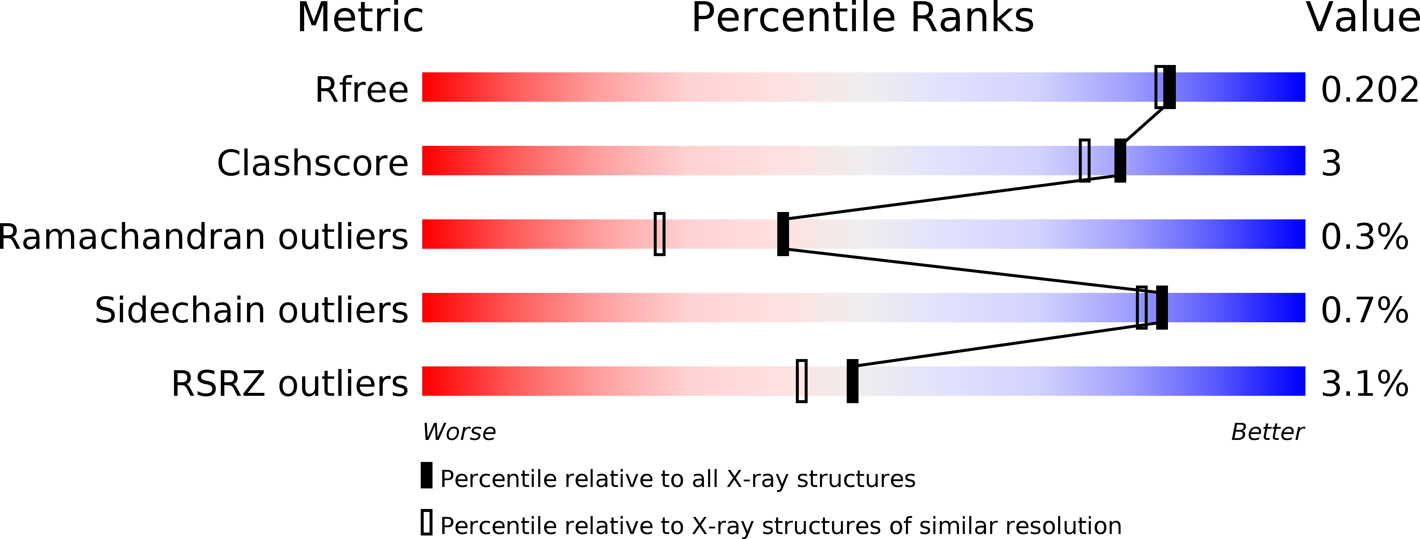
R-Value Free:
0.20
R-Value Work:
0.17
R-Value Observed:
0.17
Space Group:
H 3