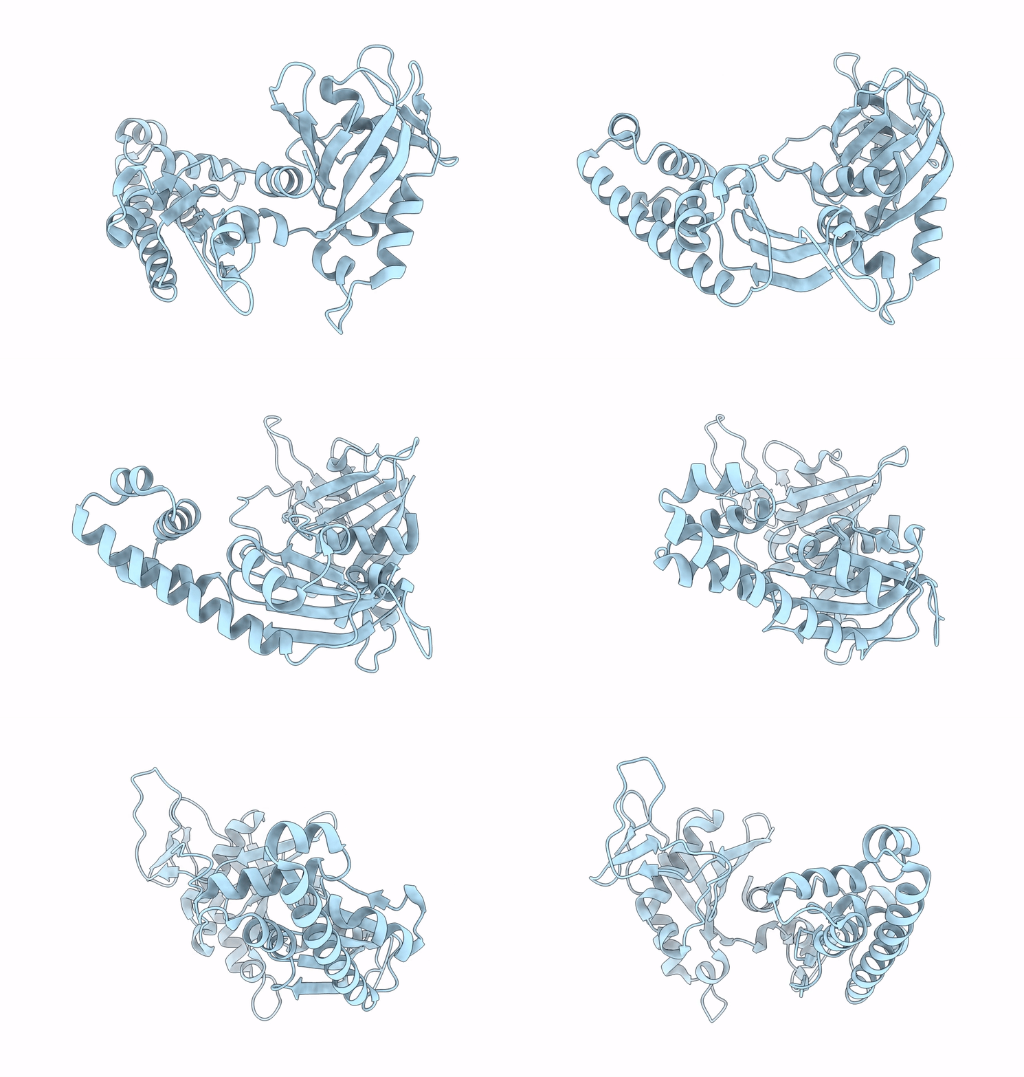
2QU4 image
Deposition Date 2007-08-03
Release Date 2007-09-18
Last Version Date 2024-02-21
Entry Detail
PDB ID:
2QU4
Title:
Model for Bacterial ParM Filament
Biological Source:
Source Organism:
Escherichia coli (Taxon ID: 562)
Method Details:
Experimental Method:
Resolution:
16.00 Å
Aggregation State:
FILAMENT
Reconstruction Method:
HELICAL
Macromolecular Entities
Polymer Type:polypeptide(L)
Molecule:Plasmid segregation protein parM
Gene (Uniprot):parM
Chain IDs:A
Chain Length:320
Number of Molecules:1
Biological Source:Escherichia coli
Ligand Molecules
Primary Citation
The structure of bacterial ParM filaments.
Nat.Struct.Mol.Biol. 14 921 926 (2007)
PMID: 17873883 DOI: 10.1038/nsmb1300

Abstact

Bacterial ParM is a homolog of eukaryotic actin and is involved in moving plasmids so that they segregate properly during cell division. Using cryo-EM and three-dimensional reconstruction, we show that ParM filaments have a different structure from F-actin, with very different subunit-subunit interfaces. These interfaces result in the helical handedness of the ParM filament being opposite to that of F-actin. Like F-actin, ParM filaments have a variable twist, and we show that this involves domain-domain rotations within the ParM subunit. The present results yield new insights into polymorphisms within F-actin, as well as the evolution of polymer families.

Legend

Protein

Chemical

Disease

Primary Citation of related structures