2QTY image
Deposition Date 2007-08-03
Release Date 2008-03-04
Last Version Date 2023-08-30
Entry Detail
PDB ID:
2QTY
Keywords:
Title:
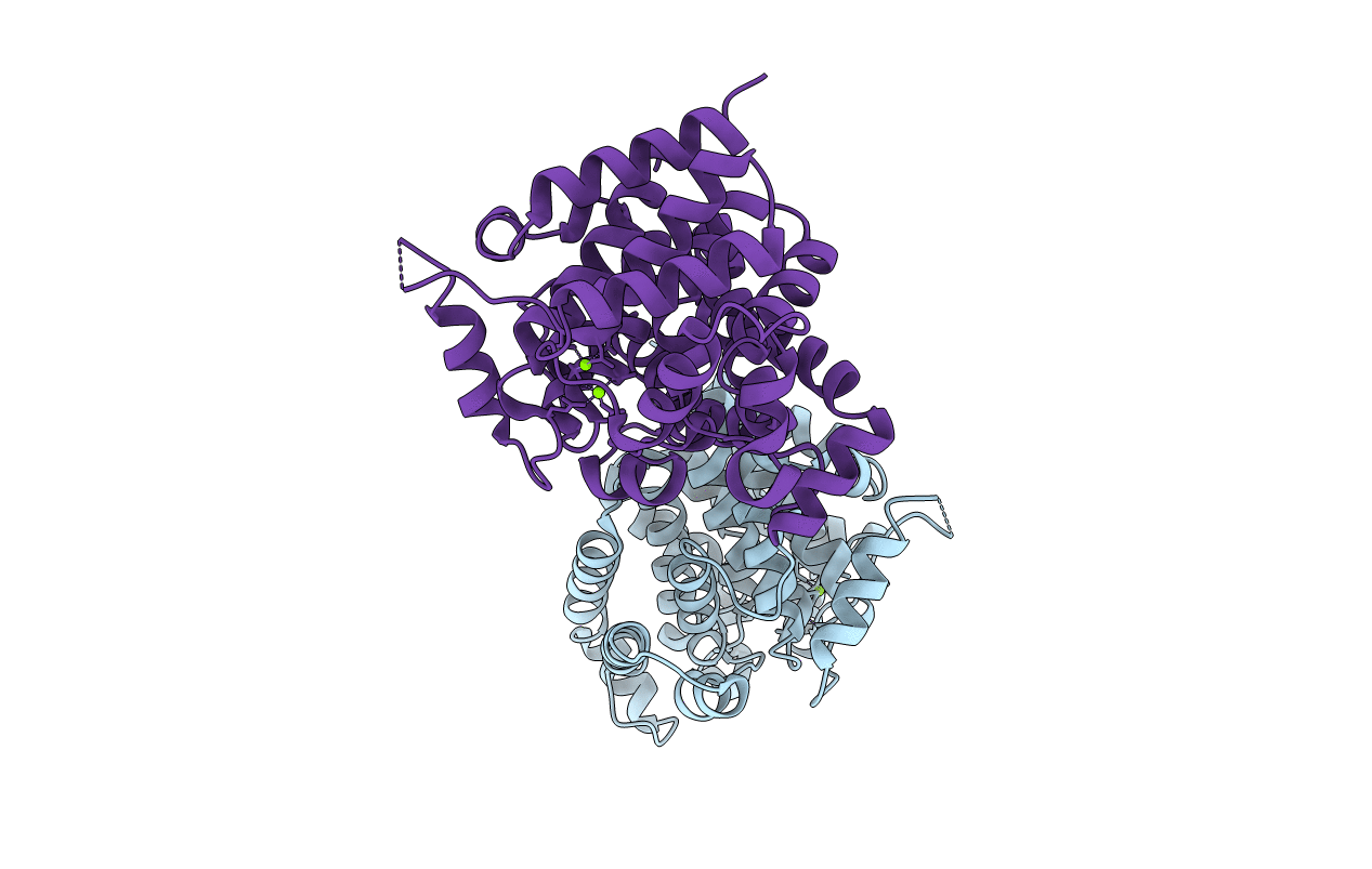
Crystal Structure of mouse ADP-ribosylhydrolase 3 (mARH3)
Biological Source:
Source Organism:
Mus musculus (Taxon ID: 10090)
Host Organism:
Method Details:
Experimental Method:
Resolution:
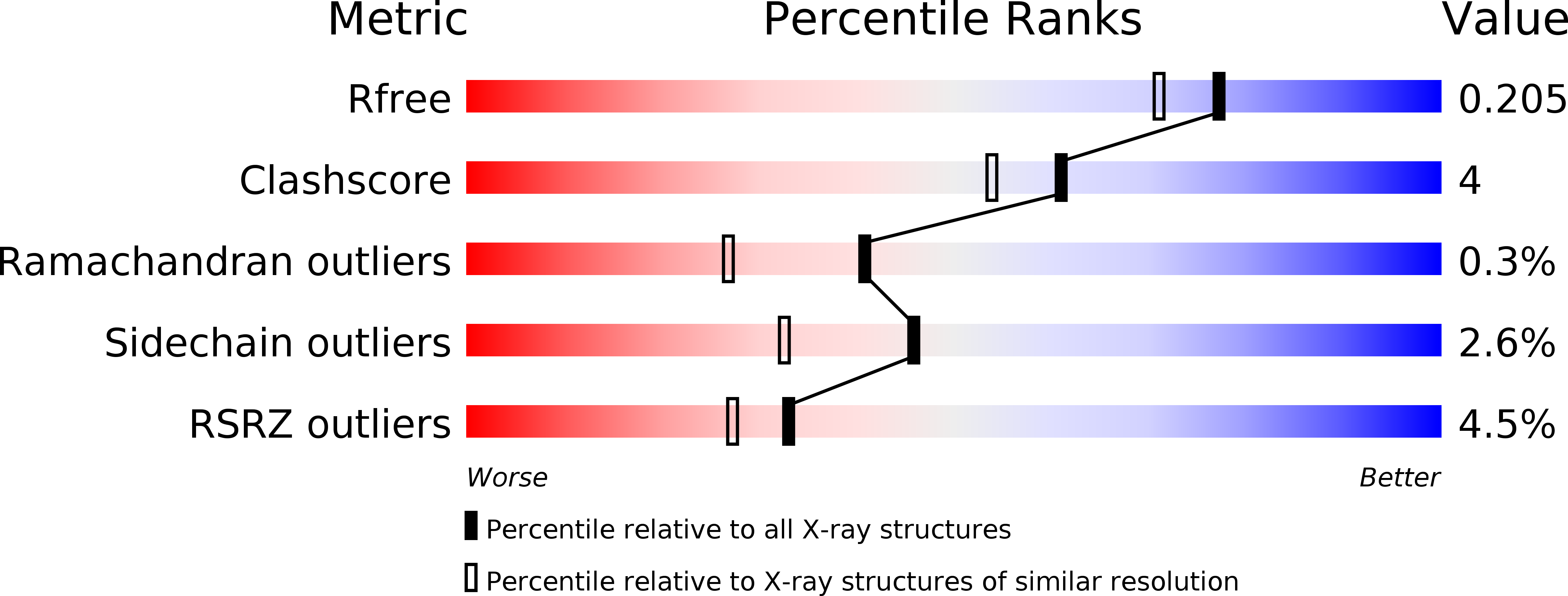
1.80 Å
R-Value Free:
0.19
R-Value Work:
0.16
R-Value Observed:
0.16
Space Group:
P 1 21 1
Macromolecular Entities
Structures with similar UniProt ID
Protein Blast
Polymer Type:polypeptide(L)
Molecule:Poly(ADP-ribose) glycohydrolase ARH3
Gene (Uniprot):Adprs
Chain IDs:A, B
Chain Length:347
Number of Molecules:2
Biological Source:Mus musculus
Ligand Molecules
Primary Citation
Structure of mouse ADP-ribosylhydrolase 3 (mARH3).
Acta Crystallogr.,Sect.F 64 156 162 (2008)
PMID: 18323597 DOI: 10.1107/S1744309108001413

Abstact

ADP-ribosylation is a reversible and covalent post-translational modification in which the attachment of ADP-ribose is catalyzed by ADP-ribosyltransferases and the removal of ADP-ribose is catalyzed by ADP-ribosylhydrolases. ADP-ribosylhydrolase 3 from mouse, consisting of 347 amino-acid residues, has been cloned, purified and crystallized. The three-dimensional structure has been resolved at a resolution of 1.8 A. The structure constitutes a compact all-alpha-helical protein with two Mg(2+) ions located in the active-site crevice. A structural comparison of mouse ADP-ribosylhydrolase 3 with its human orthologue shows a high degree of structural similarity. Furthermore, four prokaryotic proteins deposited in the PDB could be identified as being structurally related.

Legend

Protein

Chemical

Disease

Primary Citation of related structures