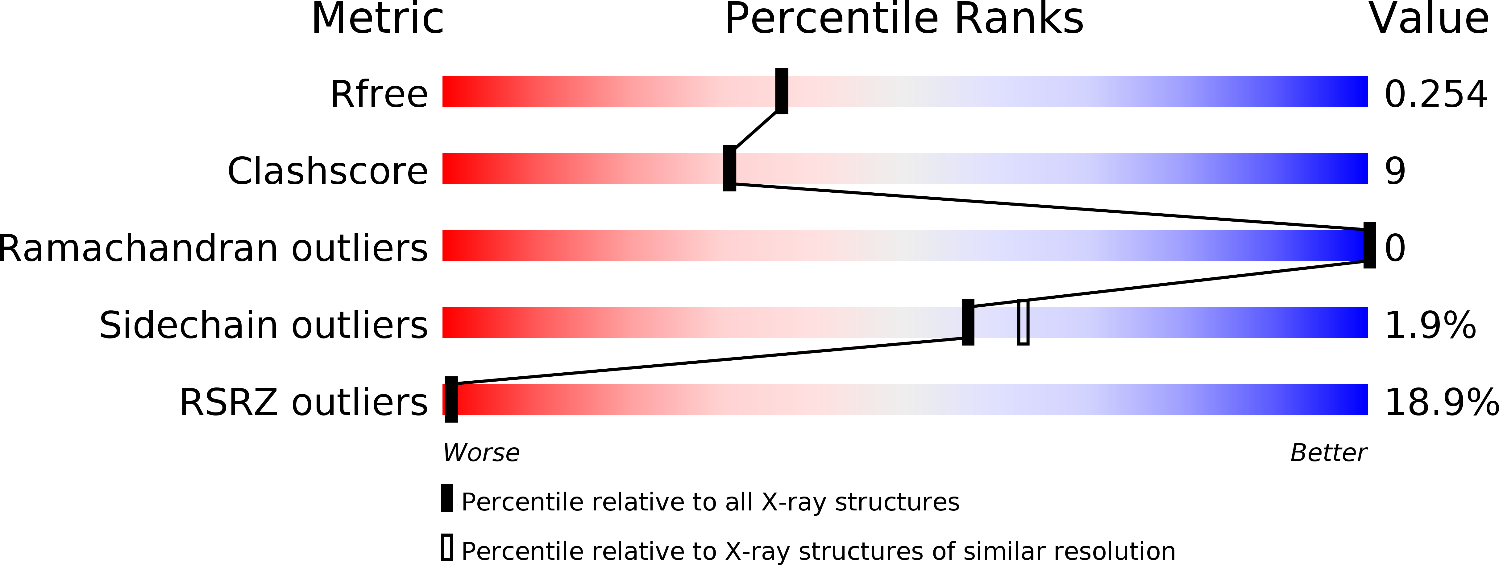
2QTP image
Deposition Date 2007-08-02
Release Date 2007-08-21
Last Version Date 2024-10-30
Entry Detail
PDB ID:
2QTP
Title:
Crystal structure of a duf1185 family protein (spo0826) from silicibacter pomeroyi dss-3 at 2.10 A resolution
Biological Source:
Source Organism:
Host Organism:
Method Details:
Experimental Method:
Resolution:
2.10 Å
R-Value Free:
0.25
R-Value Work:
0.20
R-Value Observed:
0.20
Space Group:
P 43 21 2
Macromolecular Entities
Polymer Type:polypeptide(L)
Molecule:Uncharacterized protein
Gene (Uniprot):SPO0826
Chain IDs:A
Chain Length:194
Number of Molecules:1
Biological Source:Silicibacter pomeroyi DSS-3
Modified Residue
Compound ID Chain ID Parent Comp ID Details 2D Image
MSE A MET SELENOMETHIONINE
Primary Citation

Abstact

The crystal structures of BB2672 and SPO0826 were determined to resolutions of 1.7 and 2.1 Å by single-wavelength anomalous dispersion and multiple-wavelength anomalous dispersion, respectively, using the semi-automated high-throughput pipeline of the Joint Center for Structural Genomics (JCSG) as part of the NIGMS Protein Structure Initiative (PSI). These proteins are the first structural representatives of the PF06684 (DUF1185) Pfam family. Structural analysis revealed that both structures adopt a variant of the Bacillus chorismate mutase fold (BCM). The biological unit of both proteins is a hexamer and analysis of homologs indicates that the oligomer interface residues are highly conserved. The conformation of the critical regions for oligomerization appears to be dependent on pH or salt concentration, suggesting that this protein might be subject to environmental regulation. Structural similarities to BCM and genome-context analysis suggest a function in amino-acid synthesis.

Legend

Protein

Chemical

Disease

Primary Citation of related structures