2QTK image
Deposition Date 2007-08-02
Release Date 2008-06-24
Last Version Date 2024-02-21
Entry Detail
PDB ID:
2QTK
Title:
Crystal Structure of the outer membrane protein opdK from Pseudomonas aeruginosa
Biological Source:
Source Organism:
Host Organism:
Method Details:
Experimental Method:
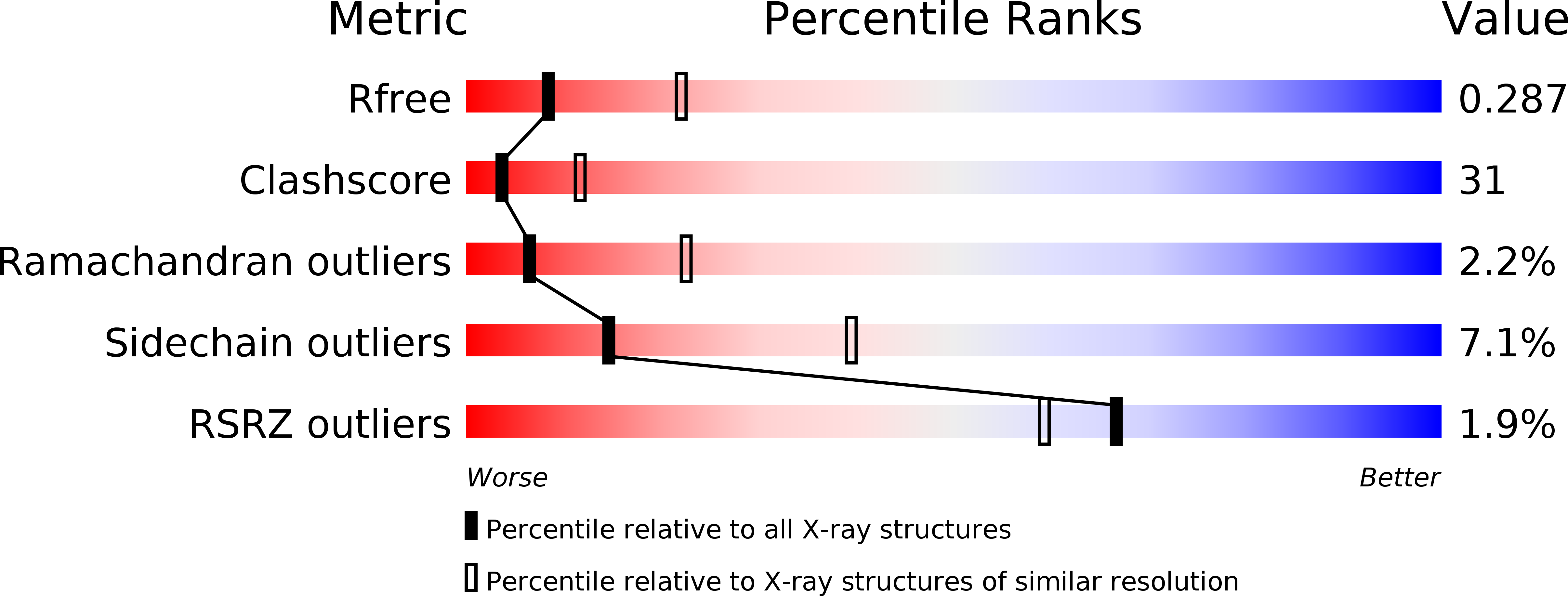
Resolution:
2.80 Å
R-Value Free:
0.29
R-Value Work:
0.23
R-Value Observed:
0.23
Space Group:
P 21 21 21
Macromolecular Entities
Structures with similar UniProt ID
Protein Blast
Polymer Type:polypeptide(L)
Molecule:Probable porin
Gene (Uniprot):opdK
Chain IDs:A, B
Chain Length:390
Number of Molecules:2
Biological Source:Pseudomonas aeruginosa
Ligand Molecules
Primary Citation
Crystal Structure of the Outer Membrane Protein OpdK from Pseudomonas aeruginosa.
Structure 16 1027 1035 (2008)
PMID: 18611376 DOI: 10.1016/j.str.2008.04.009

Abstact

In Gram-negative bacteria that do not have porins, most water-soluble and small molecules are taken up by substrate-specific channels belonging to the OprD family. We report here the X-ray crystal structure of OpdK, an OprD family member implicated in the uptake of vanillate and related small aromatic acids. The OpdK structure reveals a monomeric, 18-stranded beta barrel with a kidney-shaped central pore. The OpdK pore constriction is relatively wide for a substrate-specific channel (approximately 8 A diameter), and it is lined by a positively charged patch of arginine residues on one side and an electronegative pocket on the opposite side-features likely to be important for substrate selection. Single-channel electrical recordings of OpdK show binding of vanillate to the channel, and they suggest that OpdK forms labile trimers in the outer membrane. Comparison of the OpdK structure with that of Pseudomonas aeruginosa OprD provides the first qualitative insights into the different substrate specificities of these closely related channels.

Legend

Protein

Chemical

Disease

Primary Citation of related structures