
Deposition Date
2007-07-30
Release Date
2008-08-05
Last Version Date
2024-11-06
Entry Detail
PDB ID:
2QS4
Keywords:
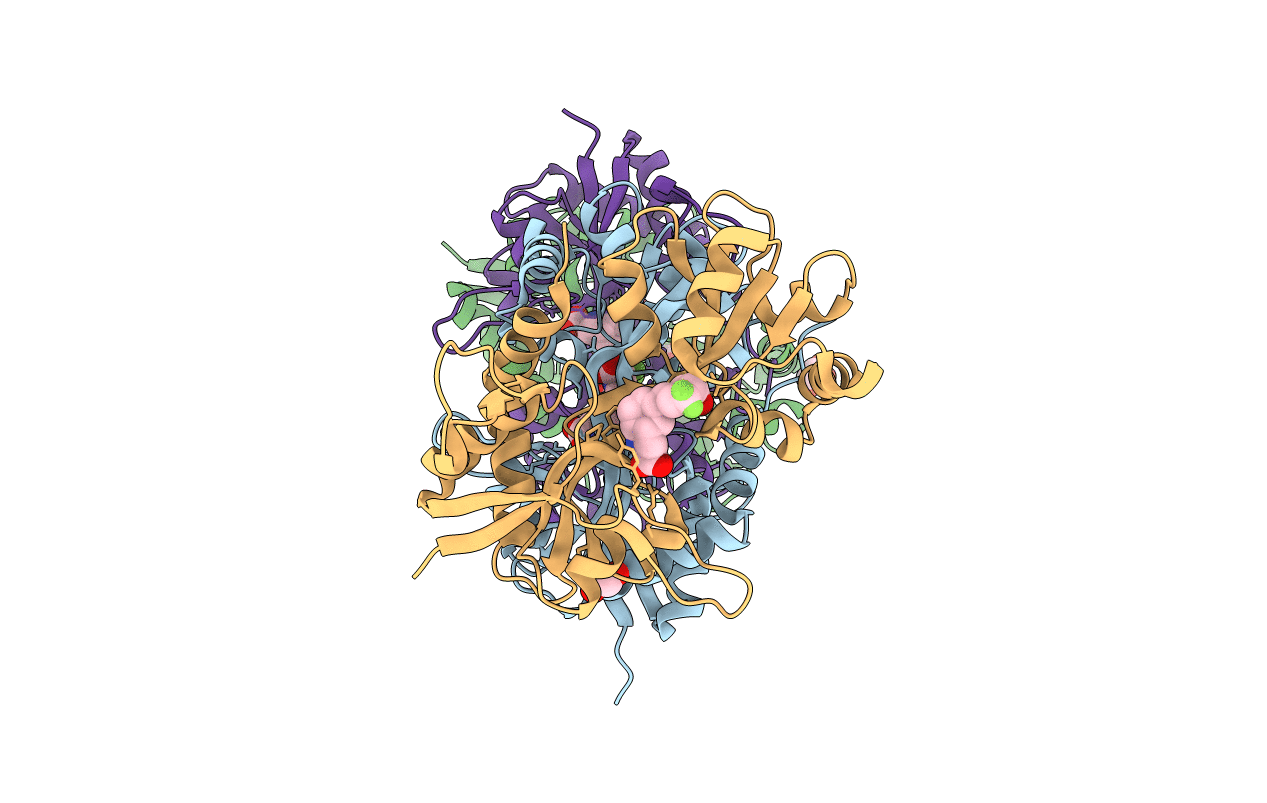
Title:
Crystal structure of the GluR5 ligand binding core dimer in complex with LY466195 at 1.58 Angstroms resolution
Biological Source:
Source Organism(s):
Rattus norvegicus (Taxon ID: 10116)
Expression System(s):
Method Details:
Experimental Method:
Resolution:
1.58 Å
R-Value Free:
0.19
R-Value Work:
0.15
R-Value Observed:
0.16
Space Group:
H 3
