Deposition Date
2007-07-30
Release Date
2007-11-06
Last Version Date
2023-08-30
Entry Detail
PDB ID:
2QRW
Keywords:
Title:
Crystal structure of Mycobacterium tuberculosis trHbO WG8F mutant
Biological Source:
Source Organism(s):
Mycobacterium tuberculosis (Taxon ID: 1773)
Expression System(s):
Method Details:
Experimental Method:
Resolution:
1.93 Å
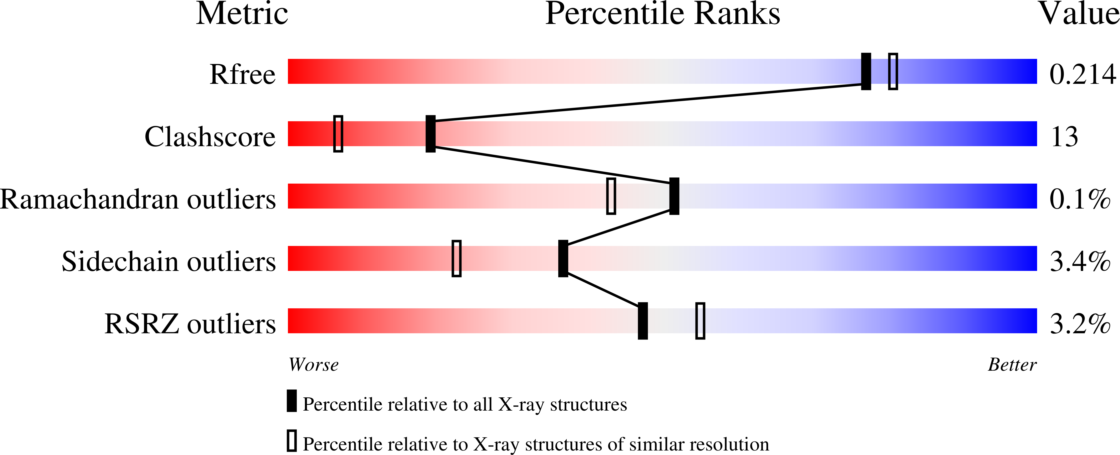
R-Value Free:
0.21
R-Value Work:
0.17
R-Value Observed:
0.17
Space Group:
I 41 2 2