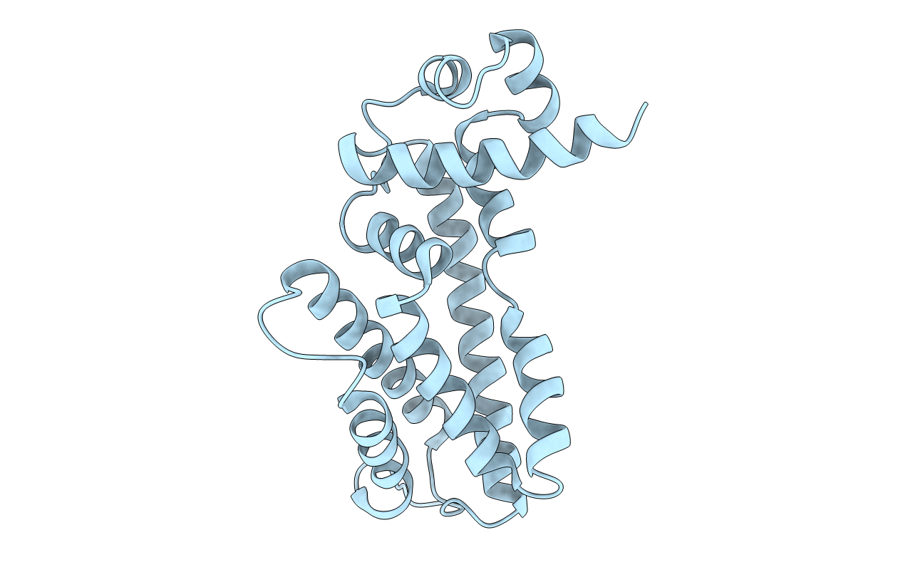
2QOP image
Deposition Date 2007-07-20
Release Date 2008-02-26
Last Version Date 2024-02-21
Entry Detail
PDB ID:
2QOP
Keywords:
Title:
Crystal structure of the transcriptional regulator AcrR from Escherichia coli
Biological Source:
Source Organism:
Host Organism:
Method Details:
Experimental Method:
Resolution:
2.55 Å
R-Value Free:
0.27
R-Value Work:
0.22
Space Group:
P 2 2 21
Macromolecular Entities
Structures with similar UniProt ID
Protein Blast
Polymer Type:polypeptide(L)
Molecule:HTH-type transcriptional regulator acrR
Gene (Uniprot):acrR
Chain IDs:A
Chain Length:215
Number of Molecules:1
Biological Source:Escherichia coli K12
Primary Citation
Crystal structure of the transcriptional regulator AcrR from Escherichia coli.
J.Mol.Biol. 374 591 603 (2007)
PMID: 17950313 DOI: 10.1016/j.jmb.2007.09.064

Abstact

The AcrAB multidrug efflux pump, which belongs to the resistance nodulation division (RND) family, recognizes and extrudes a wide range of antibiotics and chemotherapeutic agents and causes the intrinsic antibiotic resistance in Escherichia coli. The expression of AcrAB is controlled by the transcriptional regulator AcrR, whose open reading frame is located 141 bp upstream of the acrAB operon. To understand the structural basis of AcrR regulation, we have determined the crystal structure of AcrR to 2.55-A resolution, revealing a dimeric two-domain molecule with an entirely helical architecture similar to members of the TetR family of transcriptional regulators. Each monomer of AcrR forms a multientrance pocket of 350 A(3) in the ligand-binding domain. The ligand-binding pocket is surrounded with mostly hydrophobic residues. In addition, a completely buried negatively charged glutamate, expected to be critical for drug binding, is located at the center of the binding pocket. The crystal structure provides novel insight into the mechanisms of ligand binding and AcrR regulation.

Legend

Protein

Chemical

Disease

Primary Citation of related structures