
Deposition Date
2007-07-19
Release Date
2007-09-04
Last Version Date
2024-11-06
Entry Detail
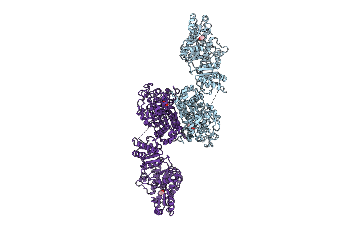
PDB ID:
2QO3
Keywords:
Title:
Crystal Structure of [KS3][AT3] didomain from module 3 of 6-deoxyerthronolide B synthase
Biological Source:
Source Organism(s):
Saccharopolyspora erythraea (Taxon ID: 1836)
Expression System(s):
Method Details:
Experimental Method:
Resolution:
2.59 Å
R-Value Free:
0.26
R-Value Work:
0.21
R-Value Observed:
0.21
Space Group:
P 1 21 1
