2QL2 image
Deposition Date 2007-07-12
Release Date 2008-11-04
Last Version Date 2023-08-30
Entry Detail
PDB ID:
2QL2
Title:
Crystal Structure of the basic-helix-loop-helix domains of the heterodimer E47/NeuroD1 bound to DNA
Biological Source:
Source Organism:
Mus musculus (Taxon ID: 10090)
(Taxon ID: )
Host Organism:
Method Details:
Experimental Method:
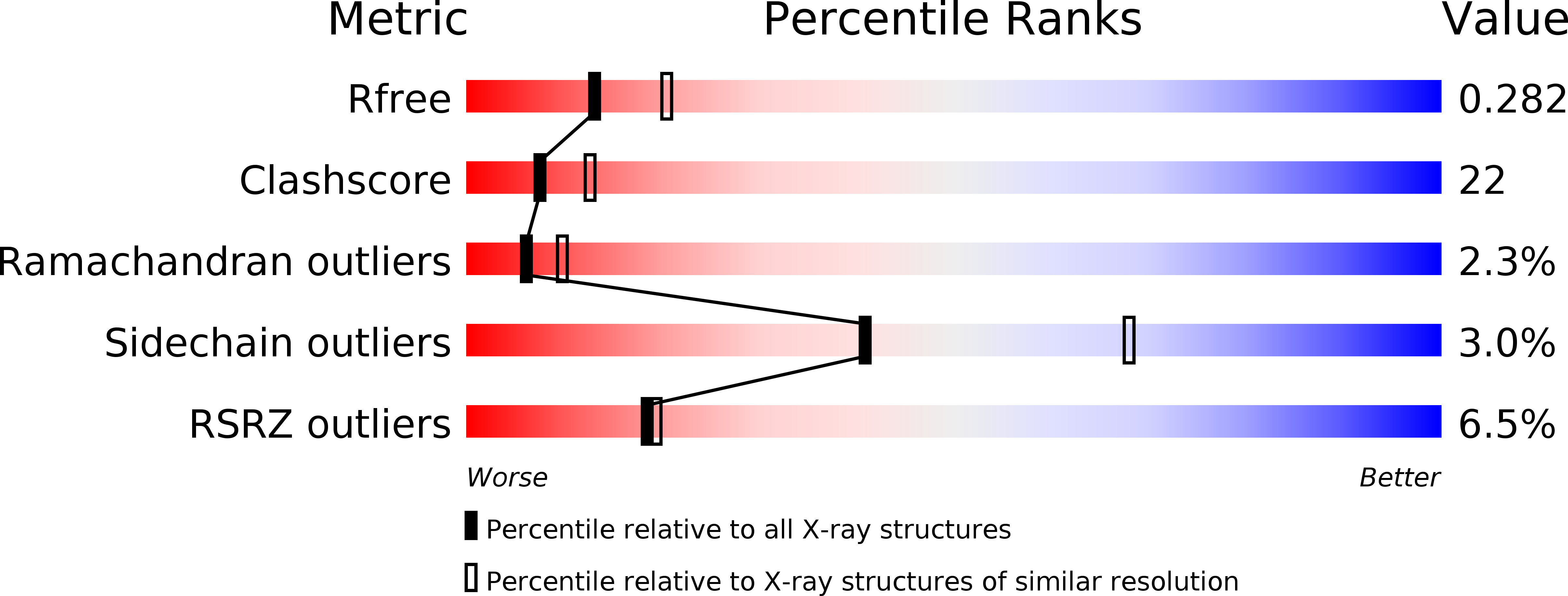
Resolution:
2.50 Å
R-Value Free:
0.28
R-Value Work:
0.24
Space Group:
P 21 21 2
Macromolecular Entities
Polymer Type:polypeptide(L)
Molecule:Transcription factor E2-alpha
Chain IDs:A, C
Chain Length:60
Number of Molecules:2
Biological Source:Mus musculus
Polymer Type:polypeptide(L)
Molecule:Neurogenic differentiation factor 1
Gene (Uniprot):Neurod1
Chain IDs:B, D
Chain Length:60
Number of Molecules:2
Biological Source:Mus musculus
Polymer Type:polydeoxyribonucleotide
Molecule:DNA (5'-D(*DTP*DAP*DGP*DGP*DCP*DCP*DAP*DTP*DCP*DTP*DGP*DGP*DTP*DCP*DCP*DT)-3')
Chain IDs:E, G
Chain Length:16
Number of Molecules:2
Biological Source:
Polymer Type:polydeoxyribonucleotide
Molecule:DNA (5'-D(*DAP*DGP*DGP*DAP*DCP*DCP*DAP*DGP*DAP*DTP*DGP*DGP*DCP*DCP*DTP*DA)-3')
Chain IDs:F, H
Chain Length:16
Number of Molecules:2
Biological Source:
Primary Citation
Crystal structure of E47-NeuroD1/beta2 bHLH domain-DNA complex: heterodimer selectivity and DNA recognition.
Biochemistry 47 218 229 (2008)
PMID: 18069799 DOI: 10.1021/bi701527r

Abstact

The ubiquitous class I basic helix-loop-helix (bHLH) factor E47 forms heterodimers with multiple tissue specific class II bHLH proteins to regulate distinct differentiation pathways. In order to define how class I- class II heterodimer partners are selected, we determined the crystal structure of the E47-NeuroD1-bHLH dimer in complex with the insulin promoter E-box sequence. Purification of the bHLH domain of E47-NeuroD1 indicates that E47 heterodimers are stable in solution. The interactions between E47 and NeuroD1 in the heterodimer are comparable to the interactions between E47 monomers in the homodimer, including hydrogen bonding, buried hydrophobic surface, and packing interactions. This is consistent with a model in which E47-NeuroD1 heterodimers are favored due to the instability of NeuroD1 homodimers. Although E47-NeuroD1 is oriented uniquely on the E-box sequence (CATCTG) within the promoter of the insulin gene, no direct contacts are observed with the central base pairs within this E-box sequence. We propose that concerted domain motions allow E47 to form specific base contacts in solution. NeuroD1 is restrained from adopting the same base contacts by an additional phosphate backbone interaction by the neurogenic-specific residue His115. Orienting E47-NeuroD1 on promoters may foster protein-protein contacts essential to initiate transcription.

Legend

Protein

Chemical

Disease

Primary Citation of related structures